


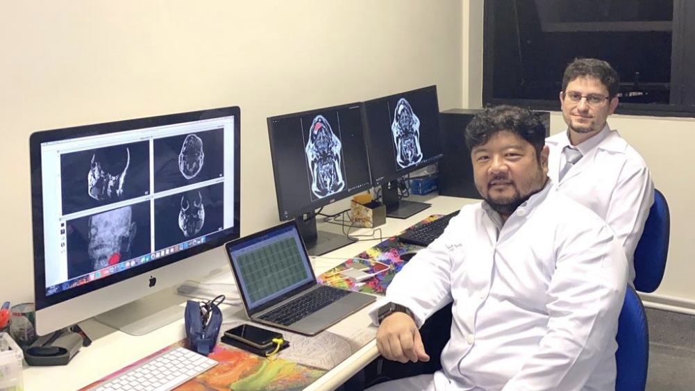
First study to identify pixel and voxel organizational parameters in MRI scans of two different (albeit similar) benign tumors may contribute to development of more accurate, less invasive diagnosis (Celso Massahiro Ogawa (UNICSUL) and João Pedro Perez Gomes (USP), first authors of the article in Scientific Reports/researchers’ achieve)
First study to identify pixel and voxel organizational parameters in MRI scans of two different (albeit similar) benign tumors may contribute to development of more accurate, less invasive diagnosis.
First study to identify pixel and voxel organizational parameters in MRI scans of two different (albeit similar) benign tumors may contribute to development of more accurate, less invasive diagnosis.
First study to identify pixel and voxel organizational parameters in MRI scans of two different (albeit similar) benign tumors may contribute to development of more accurate, less invasive diagnosis (Celso Massahiro Ogawa (UNICSUL) and João Pedro Perez Gomes (USP), first authors of the article in Scientific Reports/researchers’ achieve)
By Julia Moióli | Agência FAPESP – Ameloblastomas and odontogenic keratocysts are benign lesions of the maxillo-mandibular region with different biological characteristics. The former are often aggressive, forming a large tumor and growing into the jawbone. The latter are cysts that can be managed and treated in various ways. However, they have practically identical morphological characteristics and are extremely difficult to distinguish using conventional imaging techniques such as X-rays, MRI scans and CAT scans, making treatment hard to plan and potentially requiring additional procedures.
In search of a solution to predict the type of lesion and facilitate surgical planning, researchers at Cruzeiro do Sul University (UNICSUL), the University of São Paulo (USP) and the State University of Campinas (UNICAMP) in Brazil, in partnership with colleagues at the University of Gothenburg in Sweden and the University of Ankara in Turkey, used an image processing technique called texture analysis for the first time on these lesions. The study is described in an article published in the journal Scientific Reports.
“In three-dimensional imaging techniques, such as MRI and CAT scans, the voxels [units representative of a given volume of 3D space] and pixels are arranged differently, with distances and shades of gray that vary depending on the tissue being scanned. This information can be converted into numerical values [algorithms] to produce a purely mathematical and statistical analysis,” said André Luiz Ferreira Costa, last author of the article and a professor in UNICSUL’s program of graduate studies in dentistry.
FAPESP supported the study via two projects (2013/07559-3 and 2017/09550-4).
The study sample comprised 18 patients diagnosed with one of the lesions and undergoing treatment at the dentistry division of Hospital das Clínicas, the hospital complex run by USP’s Medical School. The diagnosis was confirmed by biopsy in all cases. Eight had ameloblastomas and ten had keratocysts. The analysis used their MRI scans, which were chosen because they are three-dimensional and do not involve radiation, so that they can be repeated.
Eleven textural parameters were measured at five different distances, for a total of 55 variables, which were processed using MATLAB scientific software. The variables “entropy” and “sum average” were found to be statistically significant. The former refers to the degree of disorder among pixels in the image of interest, while the latter refers to the average of the sums of two-pixel values in the image. Keratocysts display greater uniformity and less gray-level disorder than ameloblastomas.
“The main achievement of the study is the possibility of obtaining a definitive result more quickly by means of imaging and therefore of more suitable and safer treatment,” Costa said. “These two types of lesion are extracted differently. Keratocysts are removed and the remaining cavity is thoroughly cleaned. This is a relatively simple surgical procedure. Ameloblastomas require a larger safety margin, involving bone resection because of their aggressive behavior.”
Next steps
The results of the study are promising in so far as they show that MRI scan parameter measurement can be used to distinguish between ameloblastomas and keratocysts as an important diagnostic aid. Next steps will include studies involving other types of lesions, such as unicystic ameloblastoma, which is relatively rare, and orthokeratinized odontogenic keratocyst.
The researchers also plan to increase the size of the study sample. Because texture analysis is based on the signal intensity of pairs of pixels, the results will be better if there are more patients in the study sample and the lesions are larger.
The article “Magnetic resonance imaging texture analysis to differentiate ameloblastoma from odontogenic keratocyst” is at: doi.org/10.1038/s41598-022-20802-7.
The Agency FAPESP licenses news via Creative Commons (CC-BY-NC-ND) so that they can be republished free of charge and in a simple way by other digital or printed vehicles. Agência FAPESP must be credited as the source of the content being republished and the name of the reporter (if any) must be attributed. Using the HMTL button below allows compliance with these rules, detailed in Digital Republishing Policy FAPESP.
