


A team of biomedical specialists prepares patients for PET/CT scans (photos: Romulo Santana Osthues)
The new functionalities allow the Cancer Theranostics Innovation Center to generate dynamic, more detailed images and provide more accurate diagnoses. The device is available to the scientific community in the state of São Paulo.
The new functionalities allow the Cancer Theranostics Innovation Center to generate dynamic, more detailed images and provide more accurate diagnoses. The device is available to the scientific community in the state of São Paulo.
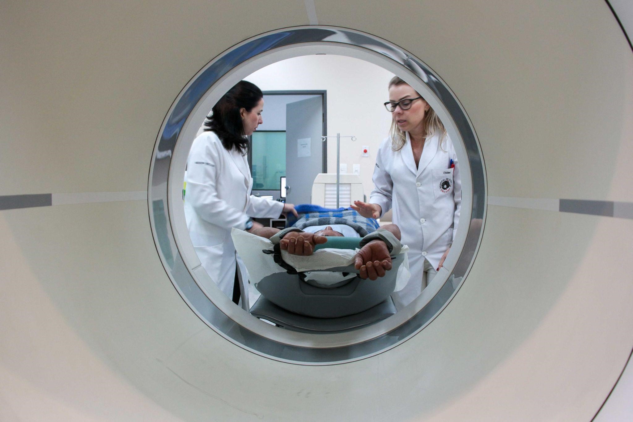
A team of biomedical specialists prepares patients for PET/CT scans (photos: Romulo Santana Osthues)
Agência FAPESP* – Positron emission tomography (PET) and computed tomography (CT) are imaging tests widely used in healthcare to analyze the inside of the body in a detailed, non-invasive way.
PET allows for the assessment of both the anatomy and metabolism of tissues and organs, identifying possible alterations. CT, on the other hand, uses X-rays and computer processing to reveal the structure of the body, generating high-resolution images in several planes.
These two tests can be performed using one piece of equipment in a combination known as a PET/CT scan. After radiopharmaceuticals are administered to the patient, the equipment generates three-dimensional images that allow for the investigation of the structures and functioning of the body as a whole.
One of these machines is used by the specialists and researchers at the Cancer Theranostics Innovation Center (CancerThera), a Research, Innovation, and Dissemination Center (RIDC) based at the State University of Campinas (UNICAMP). Thanks to funding from FAPESP, a recent upgrade of the PET/CT equipment was possible, improving its systems and incorporating new features and accessories.
The Biograph mCT PET/CT equipment, manufactured by Siemens, was installed in 2013 at the SMN-HC, the Nuclear Medicine Service of the Hospital de Clínicas (HC), the general and teaching hospital of UNICAMP. It is used by nuclear medicine and oncology teams to detect tumors and metastases, as well as by RIDC researchers carrying out preclinical and clinical studies.
In addition to testing new uses for metallopharmaceuticals and radiopharmaceuticals that are well-established in treating certain cancers, RIDC CancerThera researchers are developing new “theranostic” applications – a neologism for a model that combines therapy and diagnosis in the same approach.
“PET/CT is incorporated into medical practice and the sciences as one of the most important tests, not only in the context of cancer, but in that of many other diseases, such as inflammatory, infectious, autoimmune, metabolic, and so on,” explains Carmino Antonio de Souza, an onco-hematologist, professor at the School of Medical Sciences (FCM) at UNICAMP, principal researcher at RIDC CancerThera, and vice-president of FAPESP.
“PET/CT is a key element in theranostic studies. The new radiopharmaceuticals developed at RIDC CancerThera, labeled with positron emitters, will continue to be evaluated with this type of equipment, only now, after the upgrade, with better image quality,” says Celso Darío Ramos, a nuclear physician and professor at FCM-UNICAMP, as well as a principal researcher at RIDC CancerThera.
“We’re not the only nuclear medicine service in the country that has all these resources, but as a training and research area focused on PET, I’d say it’s one of the best equipped,” Ramos highlights.
Main new resources
In April of this year, new processing and quantification resources based on artificial intelligence (AI) were incorporated into the PET/CT, making it faster and more efficient. This also made it possible to increase the number of user licenses from three to six, allowing more users to generate and evaluate images simultaneously.
Previously, PET images were acquired in segments. Now, acquisition can be done continuously, which makes planning the scan easier and offers higher-quality visualization. This feature enables dynamic images to be generated multiple times over a previously established time interval. This makes it possible to monitor the speed at which the administered radiopharmaceutical is taken up by the tissues.
In nuclear medicine, “uptake” refers to the intensity with which tissues extract a radiopharmaceutical from the bloodstream. In the case of cancer, for example, there may be high or low uptake of the radiopharmaceutical. Its distribution throughout the body is visualized via images generated by PET/CT equipment.
“Using AI, even when there are few points of uptake in the image generated because the radiation dose has been lower, the computer can improve the appearance. This makes it easier to detect lesions,” says Ramos.
Another benefit of the included AI is that the computer can automatically identify and quantify normal and abnormal areas of the scan.
The update also includes accessories for guided biopsies. During this procedure, a needle is inserted into the exact spot on the patient’s body where the scan highlights an alteration. This makes it easier to understand what is happening at the spot where the radiopharmaceutical is picked up. The specialist performing the biopsy can follow the procedure in real time via new monitors from inside the room where the PET/CT equipment is installed.
Bárbara Juarez Amorim, a nuclear doctor, coordinator of SMN-HC-UNICAMP, and RIDC CancerThera researcher, highlights the guided biopsy feature as one of the greatest advantages of the updated PET/CT equipment because it enables the diagnosis of suspicious and undefined lesions. “Sometimes you can’t tell from the PET alone whether it’s cancer or possibly something benign, like inflammation,” she explains.
Open access and funding
The PET/CT scanner available at SMN-HC-UNICAMP is part of FAPESP’s Multiuser Equipment Program (EMU). It is accessible to the entire scientific community in the state of São Paulo. To use the scanner, please make an appointment in advance by calling +55 +19 3521-7772 or emailing soniama@hc.unicamp.br.
Those interested in using the equipment will have access to PET/CT images with a variety of radiopharmaceuticals and new visualization, processing, and guided biopsy resources for use in oncological and non-oncological research.
“FAPESP has fought hard to ensure that the high-value-added equipment it funds, which has great potential for research, education, science, etc., isn’t for the exclusive use of one researcher or a group of researchers, but rather that this equipment can be used by a variety of researchers,” Souza highlights.
The total cost of the PET/CT upgrade was approximately USD 500,000, an amount fully subsidized by FAPESP, which had already financed the purchase of the equipment in 2013. At the time, it cost USD 2.4 million.
Upgrading instead of buying new equipment made it possible to save a significant amount of money. “Currently, a new PET/CT with updated systems can cost up to USD 10 million,” says Souza. The expectation is that it will be usable in good condition for another ten years after the upgrade.
* With information from Romulo Santana Osthues of CancerThera.
The Agency FAPESP licenses news via Creative Commons (CC-BY-NC-ND) so that they can be republished free of charge and in a simple way by other digital or printed vehicles. Agência FAPESP must be credited as the source of the content being republished and the name of the reporter (if any) must be attributed. Using the HMTL button below allows compliance with these rules, detailed in Digital Republishing Policy FAPESP.
