


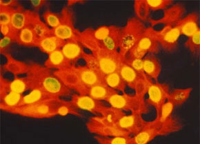
Scientists reproduce oral mucous membrane tissue from epithelial stem cells in a selected research project at FAPESP-King’s College London (photo: NYU)
Scientists reproduce oral mucous membrane tissue from epithelial stem cells in a selected research project at FAPESP-King’s College London
Scientists reproduce oral mucous membrane tissue from epithelial stem cells in a selected research project at FAPESP-King’s College London
Scientists reproduce oral mucous membrane tissue from epithelial stem cells in a selected research project at FAPESP-King’s College London (photo: NYU)
By Mônica Pileggi
Agência FAPESP – A mouth can reveal many secrets. Among them, the source of epithelial stem cells that reside in the mucous membranes, an easily-accessed tissue. Epithelial stem cells are considered to be rare and are located in specific niches. “Most existing work in the literature is about adult Mesenchymal stem cells that can be cells from bone marrow or the umbilical cord, among other places,” Andrea Mantesso, PhD explained to Agência FAPESP.
Mantesso, who is a professor of Oral Pathology at USP’s College of Dentistry, coordinates the Oral epithelial stem cells - evaluation of response to injury and self-renew capacity project alongside researcher Paul Sharpe, head of the Department of Craniofacial Development and Stem-Cell Biology at King’s College London.
The project was selected in a call for proposals released by the scientific cooperation agreement between FAPESP and King’s College London, where Sharpe and Mantesso are professors at the College of Dentistry. Signed in May 2010, the objective of the agreement is to allow for exchange of researchers between the two countries.
“The characteristics of epithelial stem cells are different from mesenchymals. It is believed, like in other tissues in the human body, that there are stem cells in the epithelium. But as there is little research on the subject, we don’t know much about them,” emphasized Mantesso.
In mesenchymal stem cells—from which it is possible to form bone, cartilage, adipose or neural tissue among others—the characterization process doesn’t happen the same way it does in epithelial cells.
One large challenge to scientists is to isolate the population of cells that have characteristics of epithelial stem cells or progenitor cells. “King’s College London’s participation is very important for this phase of the study” she pointed out.
During the first stage, done at USP, Mantesso, her doctoral student Felipe Perozzo Daltoe and Sharpe managed to isolate a population of oral mucous membrane tissue that expressed the P75NTR protein, a neurotrophine receptor considered important because it distinguishes a stem-cell rich population.
The team observed that the cells found in the experiment proliferated in greater number and more quickly than the normal ones, characteristics common to stem cells and also progenitor cells. The second part of the study will take place in London. Even though the team has already managed to rebuild an epithelium here in Brazil, Mantesso says that no denomination has been defined for the cells they found. “We need to perform more studies in vivo to confirm the potential of these epithelial cells,” she said.
This means observing one more common behavior of epithelial stem cells: the response to damage. “In this phase of the study, our intention is to study the properties of these cells in terms of migration and capacity to heal a wound. We also intend to explore their potential for engineering tissue and apply this knowledge to the regeneration and repair of teeth,” explained Sharpe.
Aside from the project funded by FAPESP, Mantesso and Sharpe studied the response to lesions in incisors by pericytes, cells that line blood vessels with other colleagues. The study’s findings were published in April’s Proceedings of the National Academy of Sciences magazine.
The study seeks to reveal the origin of mesenchyme stem cells through analysis of the pericytes. To do this, the scientists brought together a series of information common to pericytes and stem cells such as regeneration and whether both are connected to vascular activity.
Using transgenic mice, the researchers managed to see the pericytes and analyze the cells. To the group’s surprise, they weren’t the only source of mesenchymal stem cells. “During the experiments, we observed that the pericytes showed the same characteristics as trunk or progenitor cells, but they responded only on the part of the origin of these cells. And this had never been shown in the literature,” emphasized Mantesso.
According to the professor, the other cell population is as yet still unknown; however experiments indicate that they are related to vascular activity. “because these cells were more present in places rich in blood vessels,” she said.
The article Dual origin of mesenchymal stem cells contributing to organ growth and repair (doi:10.1073/pnas.1015449108), by Andrea Mantesso, Paul Sharpe and others can be ready by PNAS subscribers at: www.pnas.org/content/108/16/6503.abstract.
The Agency FAPESP licenses news via Creative Commons (CC-BY-NC-ND) so that they can be republished free of charge and in a simple way by other digital or printed vehicles. Agência FAPESP must be credited as the source of the content being republished and the name of the reporter (if any) must be attributed. Using the HMTL button below allows compliance with these rules, detailed in Digital Republishing Policy FAPESP.
