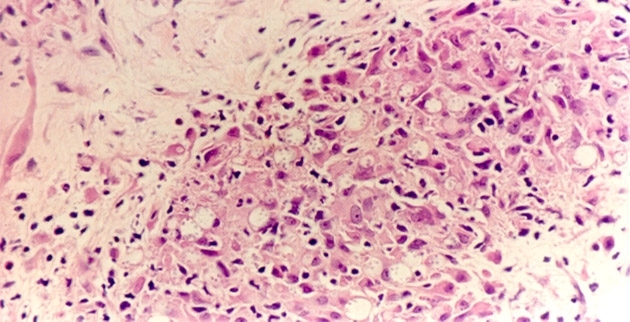

Studies carried out at the University of São Paulo and institutions in the states of Pará and Maranhão report that interaction with the host varies according to the protozoan subgenus and species (image: FMUSP)
Studies report that interaction with the host varies according to the protozoan subgenus and species.
Studies report that interaction with the host varies according to the protozoan subgenus and species.

Studies carried out at the University of São Paulo and institutions in the states of Pará and Maranhão report that interaction with the host varies according to the protozoan subgenus and species (image: FMUSP)
By Elton Alisson
Agência FAPESP – Health professionals active in the Amazon region frequently encounter patients with tegumentary (or cutaneous) leishmaniasis. These patients may present with skin lesions, which heal on their own in some who are immunologically resistant, or sores in the mucous membranes of the nose, mouth or throat, in addition to incurable infiltrated erythematous plaques and nodules throughout the body.
“For over 20 years, we have been monitoring a patient with tegumentary leishmaniasis who has lesions throughout his body that do not heal,” Carlos Eduardo Pereira Corbett, professor in the Department of Pathology and head of the Laboratory of Pathology and Infectious Diseases of the School of Medicine at the University of São Paulo (FMUSP), told Agência FAPESP. “In this case, we suggest palliative care so that the patient does not suffer as much.”
During studies carried out under a Thematic Project, a group of researchers from the institution, in collaboration with colleagues from Evandro Chagas Institute in Belém (PA) and Federal University of Maranhão (UFMA), discovered that in addition to being influenced by the genetic and immunological profile of the host, the immune response to tegumentary leishmaniasis is also determined by the species of the parasite.
Some of the most important findings from the study led by Corbett were published in the journals Archives of Dermatological Research, The Journal of the Federation of American Societies for Experimental Biology – FASEB Journal, Parasite Immunology, and Transactions of the Royal Society of Tropical Medicine and Hygiene.
“We determined that some types of cutaneous lesions found in tegumentary leishmaniasis are closely related to the species of parasite that modulate the infected patient’s immune response to develop resistance or susceptibility to the disease,” Corbett said.
The determination was made through studies carried out on patients in the states of Pará and Maranhão in areas where leishmaniasis is endemic who were diagnosed and monitored by the researchers, in some cases, for nearly 20 years.
According to Corbett, tegumentary leishmaniasis is transmitted to humans – and to wild animals such as rodents, marsupials, edentates and primates – by the bite of female phlebotominae [sand flies] (Diptera: Psychodidae) infected with the Leishmania parasite. The researchers estimate that in the past five years, nearly 30,000 cases of the disease have been reported in Brazil each year.
In the Brazilian Amazon, which has the largest variety of species of parasites in the world and the highest number of cases of infection in Brazil and in Latin America, the disease is caused by seven species of the protozoan, six from the subgenus Viannia: L. braziliensis, L. guyanensis, L. shawi, L. lainsoni, L. naiffi and L. lindenbergi, and one from the subgenus Leishmania: L. amazonensis.
When infected by one of these species of intracellular parasite, which attacks macrophage cells, the immune system of the host activates a series of defense cells and antibodies, among other mechanisms, which interact with the protozoan and determine its destruction or survival and as a result the resistance or susceptibility to the disease.
In the case of resistance to the disease, the host may develop skin lesions that heal naturally. More severe cases of infection with L. braziliensis, in which the body’s immune response to the parasite is very aggressive, may trigger sores in mucous membranes.
In cases of susceptibility, however, the host may develop, in more severe cases, incurable skin lesions all over the body, a condition known as diffuse cutaneous leishmaniasis. This occurred in a patient treated for over 20 years by researcher Fernando Silveira, of the Evandro Chagas Institute in Belém (PA), one of the co-principal investigators of the project.
“The Leishmania parasite is over 250 million years old and has adapted during this time,” the researcher said. “Understanding the mechanisms of the interaction between the parasite and the host, the focus of our research group, still presents a huge challenge,” he said.
Role of the parasite
It is known that the range of clinical and immunological responses to infection by the Leishmania parasite is mainly related to the genetic and immunological profile of the host.
The project researchers have now demonstrated that the species of parasite also plays a crucial role in determining the type of immune response.
The species of subgenus Viannia, such as L. braziliensis and L. guyanensis, induce the production of two cytokines (proteins that modulate cell function), IFN-γ and TNF-α, which cause infected macrophage cells to produce nitric oxide (NO) and eliminate the parasite. The host, in this case, develops resistance to the infection.
In contrast, the species of subgenus Leishmania, such as L. amazonensis, stimulate the production of cytokines such as interleukin-4 (IL-4), interlukin-10 (IL-10) and TGFβ1, which have the ability to suppress the function of IFN-γ and, as a result, deactivate the macrophage, leading to multiplication of the parasite and host susceptibility to the disease.
“Of course, the immunogenetic profile is important to the immune response of the host against the infection,” said Cláudia Maria de Castro Gomes, researcher at FMUSP and one of the co-principal investigators of the project. “However, we have observed through the analysis of patient’s cells and tissues that the species of the parasite also helps to polarize the response when modulating it,” she said.
Experimental infection
To determine whether the results could be observed in experiments, the researchers used model animals. In one of the studies published in the journals Parasite Immunology and Parasitology Research, the researchers infected mice with L. amazonensis and L. braziliensis.
Infection with L. amazonensis led to progression of the disease, with an increase in the size of the lesions and the parasite load in the animal. Infection with L. braziliensis, however, caused only a minor increase in the lesions between the sixth and seventh week after inoculation with the parasite, with later regression and reduction of the parasite load.
“We were able to reproduce and corroborate in the experimental model the results we observed in human infection,” said Marcia Dalastra Laurenti, professor at FMUSP as well as one of the project’s co-principal investigators.
The researchers also conducted an experimental study using five species of neotropical monkeys, Callithrix jacchus, Callithrix penicillata, Saimiri sciureus, Aotus azarae infulatus and Callimico goeldii, from the Primate Center of the Evandro Chagas Institute in Belém, Pará.
The macrophage cells of the peritoneum of the animals were infected with the species L. braziliensis and L. amazonensis, in addition to L. infantum chagasi, which causes visceral leishmaniasis that affects organs such as the liver and spleen.
The results of the experiment, published in the Journal of the São Paulo Institute of Tropical Medicine, indicated that despite being infected, the macrophage cells of the animals were able to control the infection.
“We observed that 48 hours after infection, the presence of the parasite inside the cells diminished and tended to disappear,” Laurenti said. “The cells were producing cytokines, oxygen reagents and nitrite, which are able to control growth and destroy the parasite,” she explained.
In further studies that looked for another experimental primate model susceptible to infection with Leishmania, the researchers injected the parasites L. braziliensis and L. amazonensis intradermally in the tails of capuchin monkeys (Sapajus apella).
The study results, published in late August in the journal BioMed Research International, reported that, as with the other five species of neotropical primates, the capuchin monkeys were also able to control infection with Leishmania.
The infection with L. braziliensis lasted approximately 300 days and caused lesions in the animals that healed naturally. The infection with L. amazonensis lasted less time, approximately 180 days, and also healed on its own.
“We saw that primates are good models for evolutionary studies of the localized form of the disease that culminates in natural healing and resistance,” Laurenti said. “Of the six species of primates that we studied experimentally, none presented susceptibility to visceral leishmaniasis,” she said.
Republish
The Agency FAPESP licenses news via Creative Commons (CC-BY-NC-ND) so that they can be republished free of charge and in a simple way by other digital or printed vehicles. Agência FAPESP must be credited as the source of the content being republished and the name of the reporter (if any) must be attributed. Using the HMTL button below allows compliance with these rules, detailed in Digital Republishing Policy FAPESP.


