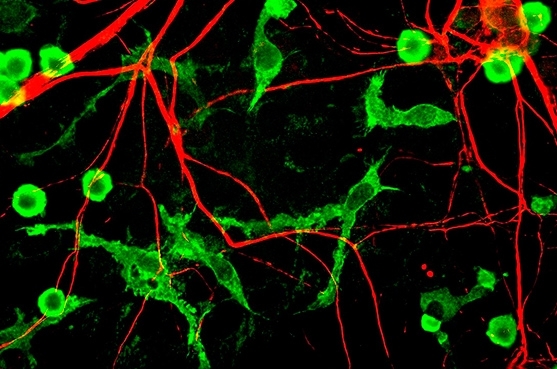

In a computer simulation, introducing a mathematical term for plasticity led to changes in the topology of a neural network, such that different synchronization patterns coexisted among the neurons (image: Wikimedia)
In a computer simulation, introducing a mathematical term for plasticity led to changes in the topology of a neural network, such that different synchronization patterns coexisted among the neurons.
In a computer simulation, introducing a mathematical term for plasticity led to changes in the topology of a neural network, such that different synchronization patterns coexisted among the neurons.

In a computer simulation, introducing a mathematical term for plasticity led to changes in the topology of a neural network, such that different synchronization patterns coexisted among the neurons (image: Wikimedia)
By José Tadeu Arantes | Agência FAPESP – With approximately 100 billion neurons, each of which has 10,000 connections to other neurons, the human brain is the most sophisticated object currently under scrutiny by contemporary science.
One aspect of the brain’s sophistication is neuroplasticity, or its ability to reorganize its synaptic pathways in response to new sensory stimuli and information, changes in environmental parameters, or damage to the previously established structure.
Synaptic plasticity can be reinforced or inhibited. This is of enormous interest not only for possible medical applications but also to help understand complex processes such as learning.
There are mathematical models that simulate the dynamics of neurons. The most famous is the Hodgkin-Huxley model, which won its creators the 1963 Nobel Prize in Physiology or Medicine. Alan Lloyd Hodgkin (1914-98) and Andrew Huxley (1917-2012), both English men, studied an axon in the squid (Loligo pealeii) to investigate how nerve impulses in neurons are initiated and propagate. They translated this physiological process into a set of non-linear differential equations to explain the underlying ionic and electrical mechanisms.
A study published recently in the journal Neural Networks used the Hodgkin-Huxley model to simulate neuroplasticity in a neural network. This study shows that an initially simple configuration can evolve into a complex topology as the neurons change their connections.
The study was conducted by Kelly Cristiane Iarosz and Iberê Luiz Caldas from the University of São Paulo (USP) in Brazil; Rafael Ribaski Borges from the Federal University of Technology – Paraná (UTFPR) in Brazil; Fernando da Silva Borges, Ewandson Luiz Lameu and Antonio Marcos Batista from the University of Ponta Grossa (UEPG) in Brazil; and Chris Antonopoulos and Murilo da Silva Baptista from the University of Aberdeen in Scotland. It received several grants from FAPESP.
“What we did was a computer simulation based on the Hodgkin-Huxley model,” Iarosz told Agência FAPESP. “We considered a set of 200 neurons integrated in a network with global coupling, meaning each neuron was connected to all the others, by means of excitatory synapses for 80% and inhibitory synapses for 20%. Without considering synaptic plasticity, there were no significant changes in the network after temporal evolution. However, when we introduced into the equations a characteristic mathematical term to represent synaptic plasticity, we found substantial modifications.”
The mathematical term is known as STDP, short for spike timing-dependent plasticity. Spikes are the electrochemical signals fired by neurons to communicate with each other.
“When STDP was inserted and we verified the evolution of the network, we observed modifications in the coupling matrix as well as considerable effects on the synchronization or desynchronization of neurons,” Iarosz said. “Insertion of the plasticity term into the model induced a new network topology, which was as non-trivial as the initial one.”
The overall process follows a pattern known as the Hebbian rule, a reference to Canadian psychologist Donald Olding Hebb (1904-1985). The Hebbian rule determines when the synapses are excited and when they are inhibited.
“Our work clearly evidenced the network’s dependence on synaptic plasticity. We started with a global coupling condition, in which each neuron was coupled with all the others, and with excitatory or inhibitory synapses. We found that the insertion of plasticity led to different diagnoses of the state of network synchronization,” Iarosz said.
Two neurons are said to be synchronized when they fire electrochemical signals at the same time. The state of network synchronization is characterized by the order parameter, a mathematical variable whose value ranges from zero, when there is no synchronization, to one, when there is total synchronization.
Plasticity induces modifications in the neural network and may reinforce the connections among certain neurons, leading to synchronization, or inhibit connections among others, leading to desynchronization.
“So the network evolves topologically as a result of plasticity. The simple topology in which each neuron is connected to all the others gives way to far more complex topologies, with sparse, moderate and dense connections coexisting,” Iarosz said.
The study’s main contribution is its description, in mathematical language, of the biological process characterized by the rearrangement of neural connections due to a wide variety of factors, including injury, degenerative disease, new experiences and learning. This dynamic malleability of the nervous system is what is called plasticity – in this case, synaptic plasticity.
“The study showed how a deterministic system can evolve in a highly complex manner,” said Caldas, who is supervising Iarosz’s postdoctoral research.
“What we did was basic science. We weren’t concerned with immediate applications,” Caldas said. “But there’s no reason why the results obtained can’t contribute to future applications. To take a hypothetical example, we know neurons are excessively synchronized in Parkinson’s disease, and if a desynchronization factor could be induced, that might possibly be a treatment strategy.”
The article “Spike timing-dependent plasticity induces non-trivial topology in the brain,” published in the journal Neural Networks, is available at http://dx.doi.org/10.1016/j.neunet.2017.01.010.

Presynaptic and postsynaptic neurons, showing the coupling region where the synapse occurs and the direction in which the electrical signal is propagated. Figure produced by the researchers and previously published in the article “Sincronização de disparos em redes neuronais com plasticidade sináptica”, Revista Brasileira de Ensino de Física, v. 37, n. 2, 2310 (2015): http://dx.doi.org/10.1590/S1806-11173721787.

Capacitive electrical circuit in which the neuronal membrane is represented by a capacitor with parallel plates, with some possible ionic channels represented as branches of the circuit. Figure produced by the researchers and previously published in the article “Sincronização de disparos em redes neuronais com plasticidade sináptica”, Revista Brasileira de Ensino de Física, v. 37, n. 2, 2310 (2015): http://dx.doi.org/10.1590/S1806-11173721787.

Illustration showing 11 vertices, with two topologies: (a) global (all vertices connected) and (b) random (few edges). Figure produced by the researchers.
Republish
The Agency FAPESP licenses news via Creative Commons (CC-BY-NC-ND) so that they can be republished free of charge and in a simple way by other digital or printed vehicles. Agência FAPESP must be credited as the source of the content being republished and the name of the reporter (if any) must be attributed. Using the HMTL button below allows compliance with these rules, detailed in Digital Republishing Policy FAPESP.


