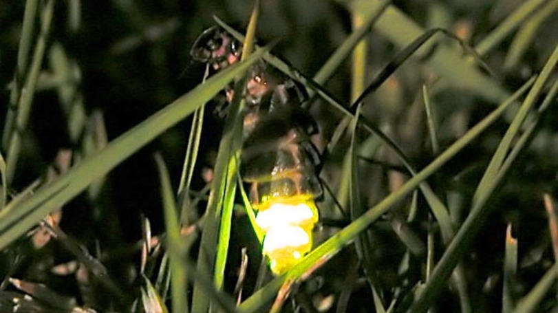

The molecular mechanisms by which luciferase emits greenish light in fireflies and reddish light in beetles have been unveiled by Brazilian researchers in collaboration with a Japanese scientist (photo: Timo Newton-Syms / Wikimedia Commons)
The molecular mechanisms by which luciferase emits greenish light in fireflies and reddish light in beetles have been unveiled by Brazilian researchers in collaboration with a Japanese scientist.
The molecular mechanisms by which luciferase emits greenish light in fireflies and reddish light in beetles have been unveiled by Brazilian researchers in collaboration with a Japanese scientist.

The molecular mechanisms by which luciferase emits greenish light in fireflies and reddish light in beetles have been unveiled by Brazilian researchers in collaboration with a Japanese scientist (photo: Timo Newton-Syms / Wikimedia Commons)
By Elton Alisson | Agência FAPESP – Enzymes known as luciferases enable fireflies and some bioluminescent species of beetle to emit cold visible light.
The same enzymes used by fireflies to emit yellow-green light at dusk, for example, explain the reddish light they emit when exposed to environments with an acidic pH, high temperatures or heavy metals. Luciferases are also responsible for the fact that beetles emit a broad range of colors regardless of the ambient pH.
In a series of research projects supported by FAPESP, scientists specializing in bioluminescence and biophotonics at the Sorocaba campus of the Federal University of São Carlos (UFSCar) in São Paulo State, Brazil, in collaboration with a colleague at the University of Electro-Communications (UEC) in Tokyo, Japan, have uncovered the molecular mechanisms that enable luciferases to emit light of different colors in bioluminescent insects.
The discoveries were described in an article published in Biochemistry, a journal of the American Chemical Society (ACS).
“Despite decades of research, the molecular mechanisms behind the changes of color in firefly and beetle bioluminescence and the sensitivity of firefly luciferase to pH, temperature and heavy metals were still unknown”, said Vadim Viviani, a professor at UFSCar and lead author of the article.
“Our study permits a better understanding of luciferase and how it can produce light of different colors,” Viviani told Agência FAPESP.
He said luciferase produces bioluminescence in fireflies and beetles by catalyzing the oxidation of luciferin, a fluorescent protein that emits light when oxidized.
The color of the light emitted varies from green to red depending on the microenvironment of the luciferase active site, Viviani explained.
The active site of an enzyme is the region in which substrate molecules bind and undergo a chemical reaction.
Research conducted in recent years at UFSCar and other universities and research institutions around the world has revealed that glutamate 311 (E311) and arginine 337 (R337), two of the 550 amino acid residues in luciferase, have opposite electric charges: E311 is negatively charged and R377 is positively charged.
Some years ago, Viviani produced a mutation in E311 to change the color of the light emitted by a Brazilian firefly’s luciferase.
A group of US researchers recently obtained the same effect by means of a mutation in R337, observing by coincidence that the residues E311 and R337 are in very close proximity in the three-dimensional structure of firefly luciferase.
“These discoveries showed that these two amino acids are important to changes in the color of the light emitted by luciferase, but exactly how they determine the color of bioluminescence has been unknown until now,” Viviani said.
The researchers and collaborators set out to answer this question using mutations in E311 and R337 that changed the electric charges of several luciferases responsible for different colors of bioluminescence, having been cloned by Viviani and his group over a period of decades.
Takashi Hirano, a professor at UEC in Japan, synthesized luciferin analogs designed to interact with specific parts of the active site in the luciferases tested.
For her Masters dissertation, Aline Simões, a graduate student in UFSCar’s biotechnology and environmental monitoring program, studied certain mutations in luciferase amino acid residues obtained from Macrolampis sp2, a firefly that inhabits Brazil’s Atlantic rainforest.
Because of their opposite electric charges , the residues interact electrostatically and close the luciferase active site. As a result, the active site becomes hydrophobic, repelling water so that the energy of the light emitted by the luciferase increases and shifts to green-blue.
Conversely, interruption of the interaction between the residues due to a change in the electric charge of one of them or in the pH of the environment in which the luciferase is located, for example, opens the active site to let water in.
“The light emitted in these situations has less energy and becomes red-shifted,” Viviani said.
One of the authors of the study, Vanessa Rezende Bevilaqua, a PhD student in UFSCar’s evolutionary genetics and molecular biology program supervised by Viviani and with a grant from FAPESP, discovered that the only luciferase that naturally emits red light, obtained from larvae of the railroad worm Phrixothrix hirtus, lacks R337 and therefore does not have a positive electric charge.
Given the absence of a positive charge, which would attract E311’s negative charge and block the active site, the region does not completely close, and the light emitted by the luciferase is red.
The results were corroborated by research using other mutations in amino acids produced by Gabriele Gabriel, also a PhD student at UFSCar, and enzyme 3D structural modeling by Frederico Arnoldi, then a student and currently a researcher at the Boticário Group Foundation for the Protection of Nature.
“These discoveries help explain how luciferase can produce light of different colors, offering the possibility of controlling these mechanisms to develop genetically engineered luciferase with the desired properties for different biotechnology applications, such as a certain color or light intensity,” Viviani said.
Biotechnology applications
Luciferase and its substrate luciferin are widely used as bioanalytical reagents and cell markers in pollution biosensors as well as in prospecting for cancer drugs and antibiotics, in addition to many other applications, he added.
Until now, their creation for biotechnology applications has been haphazard, using mutations in part of the protein to obtain a new mutant enzyme with a specific color for a targeted application. This process frequently resulted in a weak enzyme that produced little light.
“Now that we know more about the mechanisms behind the production of light, we can engineer mutations in the enzyme to change the color of the light emitted without affecting other characteristics of its bioluminescence,” Viviani said.
Viviani’s research group at UFSCar have cloned, genetically engineered and investigated more than ten different luciferases from Brazilian bioluminescent beetles, emitting light of different colors and with a range of other luminescent properties.
“We’ve cloned and investigated more insect luciferases than any other group in the world, including all the major bioluminescent families of fireflies, glow-worms and click beetles,” Viviani said.
The group’s cloning and modification of luciferase from larvae of the click beetle Pyrearinus termitilluminans, which colonizes termite mounds in the Cerrado biome, has already resulted in the development of a mammalian cell marker by a group of researchers in Japan with whom Viviani’s group collaborates.
Several other beetle luciferases cloned by the group are currently being tested for the development of biosensors and cell markers.
The article “Glu311 and Arg337 stabilize a closed active-site conformation and provide a critical catalytic base and countercation for green bioluminescence in beetle luciferases” (doi:10.1021/acs.biochem.6b00260) by Viviani et al. can be read by subscribers to Biochemistry at pubs.acs.org/doi/abs/10.1021/acs.biochem.6b00260.
Republish
The Agency FAPESP licenses news via Creative Commons (CC-BY-NC-ND) so that they can be republished free of charge and in a simple way by other digital or printed vehicles. Agência FAPESP must be credited as the source of the content being republished and the name of the reporter (if any) must be attributed. Using the HMTL button below allows compliance with these rules, detailed in Digital Republishing Policy FAPESP.


