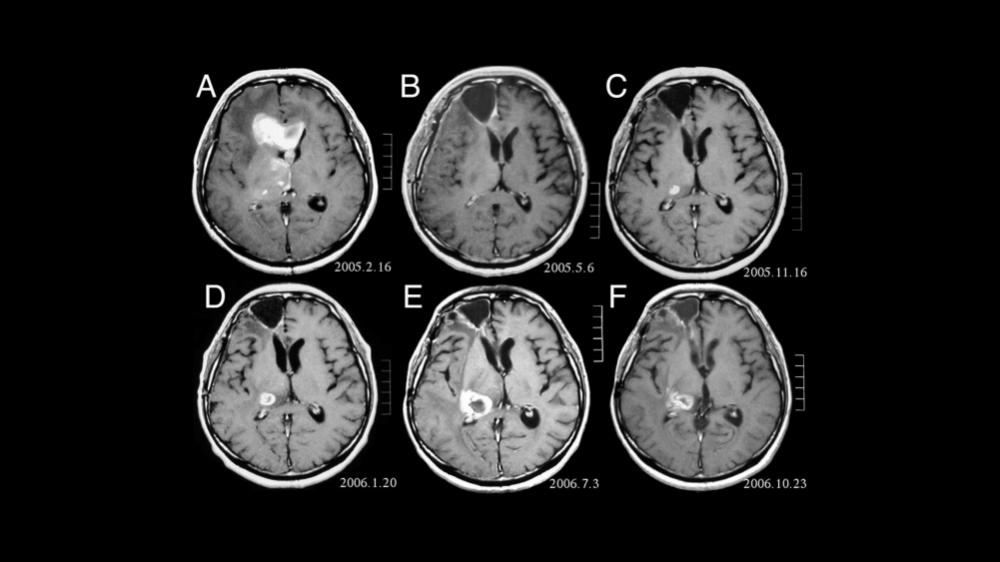

fMRI brain images – software recognizes subtle differences between structures classified as normal and those typical of people with schizophrenia (image: researcher's archive)
Software that classifies data from functional MRI already diagnoses the disease with an 80% success rate. Now, it is being used to detect alterations in brain regions associated with schizophrenia.
Software that classifies data from functional MRI already diagnoses the disease with an 80% success rate. Now, it is being used to detect alterations in brain regions associated with schizophrenia.

fMRI brain images – software recognizes subtle differences between structures classified as normal and those typical of people with schizophrenia (image: researcher's archive)
By José Tadeu Arantes | Agência FAPESP – Schizophrenia can already be diagnosed in the scientific sphere by mapping the brain with functional magnetic resonance imaging (fMRI) and data mining software. Now, researchers are working on a more detailed scrutiny of the main brain regions involved and are also attempting to detect possible reorganizations of cortical networks triggered by treatment with drugs.
This information was provided to Agência FAPESP by Francisco Aparecido Rodrigues, a researcher affiliated with the Center for Research in Mathematical Sciences Applied to Industry (CeMEAI), one of the 17 Research, Innovation and Dissemination Centers (RIDCs) supported by FAPESP.
In his capacity as a professor at the University of São Paulo’s Mathematics & Computer Science Institute (ICMC-USP) in São Carlos, São Paulo State, Brazil, Rodrigues coordinated a study on the subject in which his institution collaborated with Radboud University Nijmegen in the Netherlands. “Structure and dynamics of functional networks in child-onset schizophrenia”, reporting the initial results, was published in 2014 in the journal Clinical Neurophysiology. The study was supported by FAPESP as part of the project “Characterization, analysis, simulation and classification of complex networks”.
“In this study, which we can call an initial approach to the subject, we mapped the entire brain to detect the differences between the cortical structure classified as normal and the structure that’s typical of a person with schizophrenia,” Rodrigues said. “Now, we’re investigating several cortical regions in greater depth. These include the prefrontal cortex. The aim is to locate differences that may be even more significant. In addition, because the brain is a highly plastic organ that’s constantly changing, we want to know whether drug therapy can reconfigure linking structures and maybe lead to a definitive anatomical correction.”
The initial mapping exercise used fMRI and treated the brain as a complex network. Each node of the network represented a region of the cortex. The various regions were linked in accordance with activation during the experiment. The network was analyzed computationally using statistical descriptors and data mining methods. The analysis pointed to subtle but definite differences between the brain structures of people considered normal and those of schizophrenics.
“The brain of an individual classified as schizophrenic tends to be less organized in certain regions,” Rodrigues said. “This organizational deficit appears to be responsible for the disorders that characterize the disease in terms of sight, hearing and even smell.”
According to Rodrigues, human observers cannot distinguish between the various cortical networks even if they are specialists in the field because the networks are visually very similar, with only very minute differences in structure. “But, we used an ordinary PC and data mining software to separate the images into two distinct groups in a matter of minutes,” he said. “We extracted 54 measures from the cortical networks analyzed, and only four were of genuine relevance in terms of classifying subjects as healthy or schizophrenic.”
After separating the images into two groups, the next step was to use machine learning to teach the computer the features labeled “normal” and those assigned to schizophrenics. “From there on, the machine learned to classify the new tests and allocate them to one of the two sets with an 80% success rate,” he said.
Child-onset schizophrenia
The study focused on child-onset schizophrenia, which is particularly hard to diagnose using the conventional clinical method based on an interview, a questionnaire, and the interviewer’s subjective assessment. “This type of diagnosis is the hardest to do clinically, since the disease manifests itself in young people and children, in whom the common symptoms aren’t yet evident. However, it’s extremely important to diagnose this type of schizophrenia so that drugs can be used to prevent the disease from progressing,” Rodrigues said.
The resulting software is suitable for academic use but can be refined and made available for medical use in the future.
“The software we already have is capable of classifying data and can be used to diagnose schizophrenia in general,” Rodrigues said. “The constraints are the high cost of fMRI scans and the need for active collaboration by the person whose brain is being scanned. They can’t be sedated during the scan. They need to be wide awake and perform certain actions so the machine can detect the brain regions that are activated.”
According to the World Health Organization (WHO), schizophrenia affects more than 21 million people worldwide. It is more common among males (12 million) than females (9 million). It tends to start earlier among men. The disease typically begins in early adulthood, between the ages of 15 and 25. The average age of onset is 18 in men and 25 in women.
The software developed by the researchers can also be used to diagnose other diseases that have neural counterparts. “We’re researching its use to diagnose autism,” Rodrigues said. “It’s also possible that it could be used for early diagnosis of Alzheimer’s. This will be the medicine of the future, using computer intelligence methods to diagnose diseases that can’t easily be detected by traditional methods. Providing an accurate and fast diagnosis that’s non-invasive whenever possible is one of the major challenges facing modern medicine.”
Republish
The Agency FAPESP licenses news via Creative Commons (CC-BY-NC-ND) so that they can be republished free of charge and in a simple way by other digital or printed vehicles. Agência FAPESP must be credited as the source of the content being republished and the name of the reporter (if any) must be attributed. Using the HMTL button below allows compliance with these rules, detailed in Digital Republishing Policy FAPESP.


