


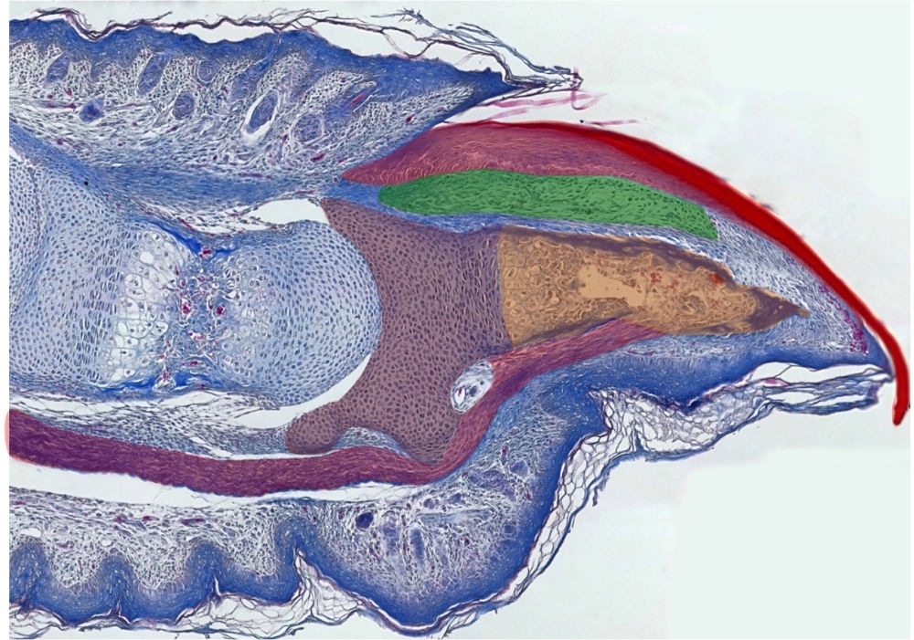
Brazilian researchers find that digit tips regrow after amputation because they consist of skin, bone and nail, three types of tissue that recover naturally when damaged (image: Lucimara Sensiate & Henrique Marques-Souza)
Brazilian researchers find that digit tips regrow after amputation because they consist of skin, bone and nail, three types of tissue that recover naturally when damaged.
Brazilian researchers find that digit tips regrow after amputation because they consist of skin, bone and nail, three types of tissue that recover naturally when damaged.
Brazilian researchers find that digit tips regrow after amputation because they consist of skin, bone and nail, three types of tissue that recover naturally when damaged (image: Lucimara Sensiate & Henrique Marques-Souza)
By André Julião in Campos do Jordão (Brazil) | Agência FAPESP – Scientists have long tried to discover the mechanisms involved in the process of digit tip regeneration (regrowth of an amputated finger or toe). This knowledge could contain the key to complete regeneration of injured or lost limbs, which is a process that occurs in salamanders.
In these amphibians, regeneration of a severed limb or tail begins with the formation of a blastema, which is a mass of undifferentiated cells capable of regrowing organs or body parts. Nothing of the kind has ever been observed in mammals.
Optimistic expectations regarding mammalian digit regeneration now appear misplaced, judging from an article published in the journal Scientific Reports by researchers affiliated with the University of Campinas (UNICAMP) in São Paulo State, Brazil. Salamanders deploy epimorphic regeneration, in which all missing parts, joints and functions of an amputated limb are restored.
Mammalian digit tip regeneration, however, involves only regrowth of nail, skin and bone.
Thus, the researchers show that the process in mammals is much simpler than expected, so tip regeneration cannot serve as a model for limb regeneration.
The study was presented on September 9, 2019, during the Third Australia-Brazil-Chile Regenerative Medicine and Developmental Biology Symposium, which was part of the 34th Annual Meeting of the Federation of Brazilian Societies for Experimental Biology (FeSBE), an event supported by FAPESP and held in Campos do Jordão, São Paulo State.
The paper was coauthored by PhD student Lucimara A. Sensiate and her doctoral advisor Henrique Marques Barbosa de Souza. Both are affiliated with UNICAMP’s Biology Institute. The study was supported by FAPESP under its Young Investigator Grants (YIG) program.
“We found that the tip is much simpler than an entire digit or limb. It lacks muscles, glands and joints and mainly comprises nail, skin and bone. These three types of tissue have a natural capacity to regenerate,” Souza said.
Strictly speaking, therefore, digit tip regrowth is not regeneration but the progression of well-known phenomena. First, the lesion caused by the amputation heals, similar to any skin wound, via migration of keratinocytes. Keratinocytes constitute 90% of the cells of the epidermis, which is the outermost layer of the skin.
When nails are cut, they regrow because they contain large proportions of stem cells that promote nail regrowth throughout the life of the organism, but bone regrowth is promoted by bone progenitor cells.
New old model
To demonstrate the regrowth phenomenon, the researchers performed experiments with mice – the murine model is the most widely used model in studying digit tip regeneration. Mice were divided in two groups and then subjected to distal and proximal amputation of the central digit of the right and left hind limbs.
Distal amputation removes approximately 25% of the digit tip and results in regrowth of a tip with morphology similar to that of an unamputated digit. Proximal amputation entails removal of 50% or more of the tip, and the wound heals without bone or nail regrowth.
To include more complex structures, the researchers performed an oblique distal amputation that removed the fat pad, a multi-tissue structure dorsally located in the digit tip and not usually affected by distal amputation. In addition to skin, the fat pad contains glands, fat and other kinds of tissue. If distally amputated digits were capable of epimorphic regeneration, they surmised, the fat pad would also regrow.
Thirty days after amputation, digits that had undergone conventional distal amputation reconstituted the claw, the distal portion of the terminal phalanx, and the skin, reproducing the original morphology, whereas obliquely amputated digits regrew all structures except the fat pad.
“This shows that structures lacking intrinsic regenerative capacity can’t regenerate after amputation,” Souza said.
Following the proximal amputations that eliminated over 50% of the digit tip, the wound healed, but the digit tip did not regrow. According to the researchers, this was because proximal amputation removed the periosteum, a membrane covering the bone surface and containing osteogenic cells that play a vital role in the growth and repair of bone tissue. Research by other groups has demonstrated the essential role played by the periosteum in the annual regeneration of deer antlers, pointing to a link between these cells and bone growth in mammals.
Cells in the periosteum are stimulated by the Wnt signaling pathway, which leads mesenchymal cells to differentiate into bone progenitor cells and to secrete a bone matrix. Previous research has shown that the presence of this signaling pathway in the nail (or claw) bed is fundamental to bone growth following distal amputation.
This signaling pathway appears to be inactive in proximal amputations, so there is no bone regrowth. When other research groups forced signaling in proximal amputations, they succeeded in reversing the phenotype, and the regenerated digit displayed bone regrowth with a similar shape to that of the original.
Studies by other groups have also added support to the theory that digital tip regeneration is basically bone regrowth by treating proximally amputated digits with beads containing bone morphogenetic proteins (BMPs), which are used in dental treatment and bone fracture healing. In these studies, the bone grew into a digital tip even without periosteum.
“In short, what needs to happen after amputation is basically bone growth stimulation. We observed the rest [wound healing] in highly similar form in distal and proximal amputations,” Souza said.
“For example, a bunion is a bony bump that forms on the joint at the base of the big toe, but no one calls it a toe because it has neither a joint nor a nail. The same applies to digit tips that regrow after amputation. They aren’t new digits; they are bone spurs.”
The researchers expect that as a result of the new findings, so-called “digit tip regeneration” will be considered only a bone growth model, and research on limb regeneration in mammals can move in new directions.
The article “Bone growth as the main determinant of mouse digit tip regeneration after amputation” (doi: 10.1038/s41598-019-45521-4) by Lucimara A. Sensiate and Henrique Marques Barbosa de Souza can be read at: www.nature.com/articles/s41598-019-45521-4.
The Agency FAPESP licenses news via Creative Commons (CC-BY-NC-ND) so that they can be republished free of charge and in a simple way by other digital or printed vehicles. Agência FAPESP must be credited as the source of the content being republished and the name of the reporter (if any) must be attributed. Using the HMTL button below allows compliance with these rules, detailed in Digital Republishing Policy FAPESP.
