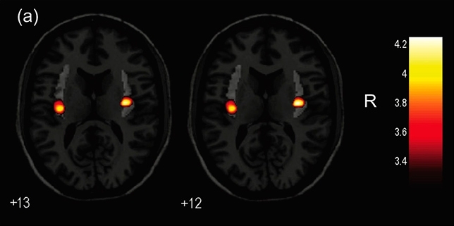

As published in Scientific Reports, research conducted in Brazil using magnetic resonance imaging points to variations in the volume of the insula, a brain region associated with body image (image: release)
As published in Scientific Reports, research conducted in Brazil using magnetic resonance imaging points to variations in the volume of the insula, a brain region associated with body image.
As published in Scientific Reports, research conducted in Brazil using magnetic resonance imaging points to variations in the volume of the insula, a brain region associated with body image.

As published in Scientific Reports, research conducted in Brazil using magnetic resonance imaging points to variations in the volume of the insula, a brain region associated with body image (image: release)
By Maria Fernanda Ziegler | Agência FAPESP – Researchers at the University of São Paulo’s Medical School (FM-USP) in Brazil used magnetic resonance imaging (MRI) to complete the first study conducted in Latin America to investigate brain volumes in transgender individuals.
They performed a structural analysis in search of differences in gray and white matter volume based on MRI scans of the brains of 80 individuals between 18 and 49 years of age.
The participants, who were all volunteers, were divided into four groups of 20 each: cisgender women, cisgender men, transgender women who had never used hormones, and transgender women who had used hormones for at least a year.
“Cisgender usually denotes people whose sense of personal identity and gender corresponds with their birth sex, while transgender, on the whole, refers to people whose sense of their own gender differs from what would be expected based on the sex characteristics with which they were born,” said Giancarlo Spizzirri, first author of a study published in Scientific Reports. The study was supported by FAPESP.
The results showed that the volume of the brain region called the insula was smaller in both hemispheres for both groups of transgender women. The insula is important to body image, among other things. The size of the insula was not smaller in transgender women than in cisgender men, but its volume was reduced in transgender women compared to cisgender women.
“It’s important to recall that there’s no such thing as a typically female or male brain. There are slight structural differences, which are far more subtle than the difference in genitals, for example. Brain structures vary greatly among individuals,” Spizzirri said.
The researchers also stressed that reduced gray matter volume in a brain region does not necessarily mean the region in question contains fewer nerve cells.
“The various gray matter brain regions contain a mass of synapses and nerve endings (called neuropils) that can change volume dynamically. For example, at any time during one’s life, a brain region’s density may increase owing to more activity, leading to a subtle rise in the volume of local gray matter,” said Professor Geraldo Busatto, who heads the Psychiatric Neuroimaging Laboratory (LIM21) at FM-USP’s general and teaching hospital (Hospital das Clínicas) and was an associate researcher in the study.
Previous studies have found that sexual differentiation of the brain in transgender individuals does not accompany differentiation in the rest of the body. “The first studies with images of the brains of transgender individuals looked for structures, regions or even functional parts of the brain that most resembled those of people of the gender with which those individuals identified. We found that trans people have characteristics that bring them closer to the gender with which they identify and [that] their brains have particularities, suggesting that the differences begin to occur during gestation,” Spizzirri said.
New research field
Another important contribution of the study is that it shows transgender “doesn’t just refer to different kinds of behavior that people develop”, according to Carmita Abdo, coordinator of the Sexuality Research Program (ProSex) at the Psychiatry Institute of Hospital das Clínicas and principal investigator of the study.
“We observed specificities in the brains of trans individuals, an important finding in light of the idea of gender ideology. The evidence is building up that it’s not a matter of ideology. Our own research based on MRI scans points to a detectable structural basis,” Abdo said.
The insula plays a key role in body image and self-awareness. Autonomic control, homeostatic information and visceral sensations are processed within the central nervous system by the insula, for example.
“It would be simplistic to make a direct link with transgender, but the detection of a difference in the insula is relevant since trans people have many issues relating to their perception of their own body because they don’t identify with the sex assigned at birth, and in addition, they unfortunately suffer discrimination and persecution,” Busatto said.
The finding cannot be seen as indicating specificity, however. “The insula is a region with multiple elements,” he stressed.
Because the insula volume differed between both groups of trans women, the authors hypothesized that this finding might to be a characteristic of trans women. Another conclusion of the study was that reduced insula volumes could not be explained by hormone treatment.
“We hope this study will be replicated with larger samples, but right now, it can be said that the hypothesis of transgender development is supported and merits investigation,” Abdo said.
The study is expected to stimulate interest in research on the brain structure of transgender people. The use of MRI scans in structural psychiatric and neurological research has increased in recent decades, thanks mainly to more advanced technology and data analysis, but few such studies have focused on transgender people.
“It’s a new research field, and this study puts Brazil among the pioneers,” Abdo said. “On the other hand, since 1997, the Federal Board of Medicine in Brazil has had guidelines on how to work with the needs of transgender individuals in clinical and surgical practice. These guidelines are periodically updated and adapted in response to new knowledge.”
The researchers plan to conduct more studies. A key interest is determining the stage of development in which differences occur. “Having detected these differences, we should try to find out when they begin to emerge. Among other points, it would be interesting to study [the] brain scans of children and young adults with transgender characteristics and compare them with the scans of adult trans women.”
The article “Gray and white matter volumes either in treatment-naïve or hormone-treated transgender women: a voxel-based morphometry study” (doi: 10.1038/s41598-017-17563-z) by Giancarlo Spizzirri, Fábio Luis Souza Duran, Tiffany Moukbel Chaim-Avancini, Mauricio Henriques Serpa, Mikael Cavallet, Carla Maria Abreu Pereira, Pedro Paim Santos, Paula Squarzoni, Naomi Antunes da Costa, Geraldo F. Busatto and Carmita Helena Najjar Abdo can be read in Scientific Reports at nature.com/articles/s41598-017-17563-z.
Republish
The Agency FAPESP licenses news via Creative Commons (CC-BY-NC-ND) so that they can be republished free of charge and in a simple way by other digital or printed vehicles. Agência FAPESP must be credited as the source of the content being republished and the name of the reporter (if any) must be attributed. Using the HMTL button below allows compliance with these rules, detailed in Digital Republishing Policy FAPESP.


