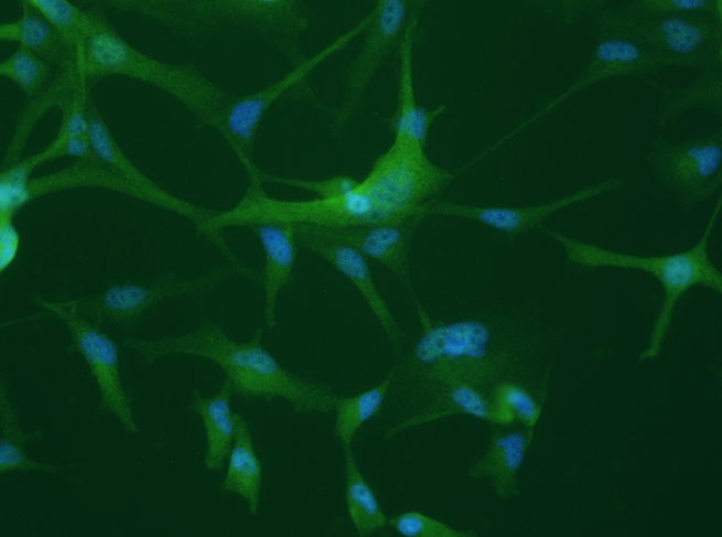

The differential expression of hnRNPs may lead to dysfunction of oligodendrocytes, glial cells that produce myelin and are important for neuronal activity (image: confocal microscope image of cultured human oligodendrocytes, by Daniel Martins-de-Souza)
The differential expression of hnRNPs may lead to dysfunction of oligodendrocytes, glial cells that produce myelin and are important for neuronal activity.
The differential expression of hnRNPs may lead to dysfunction of oligodendrocytes, glial cells that produce myelin and are important for neuronal activity.

The differential expression of hnRNPs may lead to dysfunction of oligodendrocytes, glial cells that produce myelin and are important for neuronal activity (image: confocal microscope image of cultured human oligodendrocytes, by Daniel Martins-de-Souza)
By Karina Toledo | Agência FAPESP – In the human organism, a single gene can give rise to different proteins according to the needs of the moment and in response to stimuli from the environment.
For this purpose, messenger RNA – a molecule that is transcribed from the gene and then translated into a protein – undergoes an editing or splicing process in the cell nucleus.
This editing is performed by a protein complex called the spliceosome and consists of removing introns (portions that do not contain information for the production of proteins) from the messenger RNA precursor molecule and rearranging exons (protein-encoding portions of the genetic code). The protein formed at the end of the process depends on how the spliceosome assembles the exons.
A Brazilian study supported by FAPESP and recently published in the journal Molecular Neuropsychiatry suggests that this cellular machinery for processing messenger RNA may be altered in patients with schizophrenia.
According to the authors, defects in the spliceosome could be the origin of many of the brain alterations found in patients with schizophrenia.
“A change in the system for processing messenger RNA could impair the expression of a great many proteins that play a key role in important biological processes, such as nucleic acid metabolism, creating a cascade effect. But this is something that needs to be confirmed in future research,” said Daniel Martins-de-Souza, a professor in the Biology Institute of the University of Campinas (IB-UNICAMP), São Paulo State, and principal investigator for the project.
The hypothesis presented by Martins-de-Souza’s group is based on an analysis of postmortem brain tissue from 12 patients with schizophrenia and from eight people without mental disease (control group). The study focused on two brain regions that previous research has shown to be morphologically and functionally altered in patients with schizophrenia: the anterior temporal lobe and the corpus callosum.
“The anterior temporal lobe is involved in auditory and visual processing, so it’s closely linked to symptoms such as psychosis and hallucinations. The corpus callosum is the brain region that contains the most glial cells [such as astrocytes, microglia and oligodendrocytes]. In previous research, we showed that patients with schizophrenia display oligodendrocyte dysfunction,” Martins-de-Souza said.
He explained that oligodendrocytes are the cells responsible for producing myelin, a fatty substance required for neurons to exchange information. Imaging studies performed in the 2000s showed that the brains of patients with schizophrenia contain fewer oligodendrocytes than the brains of healthy people. Around 2005, Martins-de-Souza’s group found that the expression of some proteins produced by oligodendrocytes – especially those belonging to the heterogeneous nuclear ribonucleoprotein (hnRNP) family – was also altered in these patients.
“Subsequent studies performed by other groups on the basis of our findings showed in animal and cellular models that alterations in hnRNPs do indeed interfere in the neuron myelination process and may impair cerebral connectivity. For this reason, we decided to study the role of these nuclear proteins in schizophrenia in greater depth,” Martins-de-Souza said.
Nuclear proteins
With the aid of a mass spectrometer and FAPESP’s support, the researchers mapped all the proteins (the proteome) found in the nuclei of the cells in the two brain regions selected for analysis, excluding proteins found in other organelles and in the cytoplasm.
“The total proteome of these cells had already been analyzed in previous research. However, given the complexity of this kind of analysis, it had not been possible to evaluate the differences in the expression of less abundant proteins. That’s why we decided to focus on the nuclear proteome,” said Verônica Saia-Cereda, a PhD student at IB-UNICAMP and the first author of the article.
When the researchers compared the results for the control group with the results for the patients with schizophrenia, they identified the molecules with altered expression in the pathological condition.
In the corpus callosum, they found 119 differentially expressed proteins, 24 of which were nuclear proteins. According to Saia-Cereda, most are involved in calcium-mediated cellular signaling, which is important both to the metabolism of mitochondria (organelles that produce energy for the cell) and to the removal of surplus amounts of the neurotransmitter dopamine from the synaptic vesicle (where information exchange between neurons occurs).
“Alterations in dopamine levels in the brain are associated with the most characteristic symptoms of schizophrenia, such as delirium and hallucinations,” Saia-Cereda said.
In the anterior temporal lobe, they found 224 differentially expressed proteins, and 76 were nuclear proteins. Eight of these nuclear proteins are involved in the functioning of the spliceosome.
“Among these eight are the hnRNPs, which play a key role both in the spliceosome and in oligodendrocyte functions. Therefore, this may be the genesis of the myelination dysfunctions associated with schizophrenia,” Martins-de-Souza said. “Our study is the first to link the spliceosome with the disease.”
According to Saia-Cereda, malfunctioning of the messenger RNA processing machinery may mean that certain proteins are not translated correctly and that their expression is altered throughout the organism, with currently unknown consequences. “How this relates to schizophrenia isn’t well understood. More research is required to find out,” he said.
Developments
In another study that is in progress with support from FAPESP, IB-UNICAMP postdoctoral student Mariana Fioramonte is working to identify the proteins that partner with hnRNPs to process messenger RNA.
Martins-de-Souza, her supervisor for the project, explained that the aim is to determine whether the proteins that associate with hnRNPs for this function are different in patients with schizophrenia and people without mental disorders.
“The next step will be to try to modulate the expression of these molecules in the laboratory and observe how the spliceosome functions when some of them are inhibited,” he said. “The idea is to try to find the cause of this dysregulation of the spliceosome. Depending on the results, it’s possible that some of these proteins can be tested as therapeutic targets.”
According to the authors of the article, schizophrenia affects approximately 1% of the world population and is the main cause of psychiatric incapacitation. Despite its high prevalence and the severity of its symptoms, little is known about the biochemical mechanisms involved in its development or progression. This kind of knowledge, they stress, is necessary to improve its diagnosis and treatment.
The article “The nuclear proteome of white and gray matter from schizophrenia postmortem brains” can be retrieved from karger.com/Article/Abstract/477299.
Republish
The Agency FAPESP licenses news via Creative Commons (CC-BY-NC-ND) so that they can be republished free of charge and in a simple way by other digital or printed vehicles. Agência FAPESP must be credited as the source of the content being republished and the name of the reporter (if any) must be attributed. Using the HMTL button below allows compliance with these rules, detailed in Digital Republishing Policy FAPESP.


