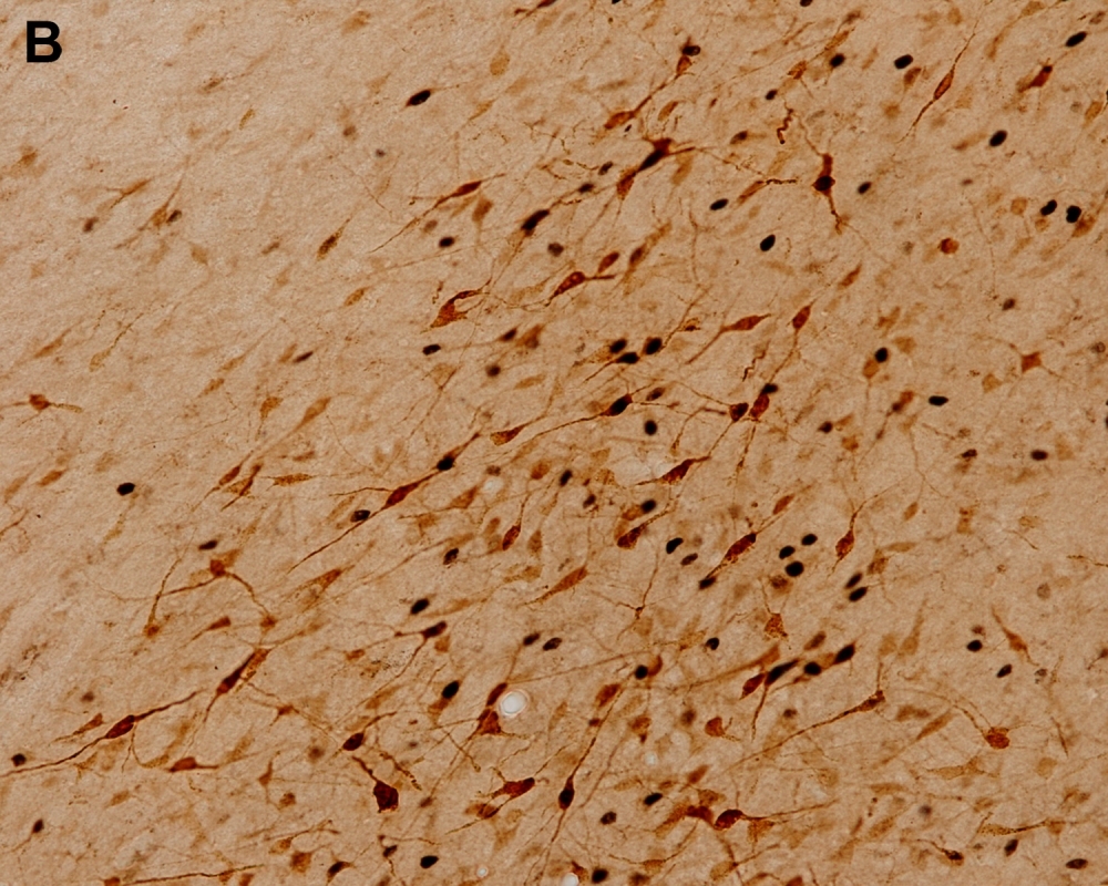

Microscope image showing active neurons in a region of the hypothalamus during social defense behavior (image: release)
Identifying the neural circuits involved in the organization of defensive responses to different dangerous situations is the goal pursued by a group of Brazilian researchers.
Identifying the neural circuits involved in the organization of defensive responses to different dangerous situations is the goal pursued by a group of Brazilian researchers.

Microscope image showing active neurons in a region of the hypothalamus during social defense behavior (image: release)
By Karina Toledo, in Rio de Janeiro
Agência FAPESP – Different dangerous situations require different defensive responses, suggesting that information reaching the brain from the outside environment must be processed through different neural circuits.
Identifying these circuits and understanding how varying responses to fear and stress are organized in the brain is the goal pursued by a group of researchers at the University of São Paulo’s Biomedical Science Institute (ICB-USP) in Brazil, via a Thematic Project supported by FAPESP and led by Newton Sabino Canteras, Full Professor at the institute.
Some of the results were presented by researcher Simone Motta during the Ninth World Congress of the International Brain Research Organization (IBRO 2015) held in Rio de Janeiro on July 7-11.
“Understanding how the brain perceives and reacts to fear can help us understand several neurological disorders,” Motta told Agência FAPESP. “The social defense model, when an individual fears a conspecific, i.e., another member of the same species, can help us understand depression, for example.”
Motta’s PhD research included investigations in animal models of the neural circuits involved in social defense behavior. The experiments consisted of placing a male “intruder” rat in a chamber previously inhabited by a male and female of the same species.
“The female was removed and another male put in its place,” Motta explained. “The older resident displayed aggressive territorial behavior, while the intruder displayed submissive behavior. This social defeat is one of the most stressful situations possible for a rat. It loses the status it enjoyed in its previous group, and its behavioral response changes drastically after this model, causing alterations similar to depression.”
Research by the ICB-USP group showed for the first time that fear of conspecifics and fear of predators activate different neural circuits. The findings were published in 2009 in the journal Proceedings of the National Academy of Sciences (PNAS) and changed the paradigm for research in this area (read more at http://revistapesquisa.fapesp.br/en/2010/05/01/the-paths-of-fear).
“It’s important that these neural pathways are different because then the response to each situation can also be appropriately different. The response to a predator depends on distance. If the predator is very close, the only chance of surviving is attack. If it’s far enough away to allow the victim to escape, the response will be flight. If the distance is shorter but still sufficient for it not to be seen, it will freeze. In social defense, however, the intruder shows its belly and raises its front paws like a boxer to ward off biting by the resident,” Motta said.
The group showed that an area of the brain called the dorsal premammillary nucleus of the hypothalamus was highly active during this social defense behavior. They identified the source of the signals received by the hypothalamus and how this brain region affected the animal’s behavior.
“In one of the experiments, we induced a lesion in the dorsal premammillary nucleus by injecting neurotoxins and observed that rats lost many social defense responses. They no longer behaved submissively and remained calmer in response to handling by researchers, apparently not suffering from stress. After attack by the resident, there was no change in the release of corticosterone, the animal equivalent of the human stress hormone cortisol,” Motta said.
With the aim of showing that this region of the hypothalamus is not involved in all types of stress but only in responses to social stress, the group used the so-called immobilization model.
“This experiment is frequently used in research on stress,” Motta explained. “The animal is placed inside a tube so that it’s unable to move. The model causes massive activation of the hypothalamic paraventricular nucleus, which is known to be related to stress. While inside the tube and until shortly after removal, the animal is highly agitated and difficult to handle.”
To the surprise of Motta and her collaborators, rats with induced dorsal premammillary lesions behaved quite differently when immobilized in this manner, remaining as calm as if nothing had happened. Subsequent tests also showed scant activation of neurons in the dorsal premammillary nucleus in this model.
“We then set out to see if the immobilization and social defense models had anything in common. Our conclusion was that it was entrapment: in both situations, the rat feels threatened and trapped, without means of escape,” Motta said.
The results of this study were published in July in the journal Physiology & Behavior.
Neurons switched on and off
According to Motta, the dorsal premammillary nucleus is activated in situations acutely important to rats, such as genuine emergencies, but depending on the situation different regions of the nucleus organize the defensive response. Each of these subregions has a different projection in the brain.
To understand the factors that influence the brain’s processing of defensive responses in greater depth, the ICB-USP research group has used genetic methods that temporarily activate or deactivate neurons in the region of interest, rather than causing permanent lesions, as in previous experiments.
One of the techniques consists of inserting the gene responsible for expressing an algal protein called opsin, which is photosensitive, into a rat’s neural cells with the aid of a modified virus. An optical fiber coupled to a laser is implanted in the brain, and a light is switched on to activate or inhibit neurons in a temporary and highly selective manner.
Another method consists of using a modified virus to insert into neurons receptor genes designed in the laboratory to respond exclusively to a specific drug created for this purpose, without any effect on other parts of the animal’s organism.
“We inject the drug, which for a few hours inhibits or activates the neurons in the region we’re studying,” Motta said. “About half an hour after the injection, we place the animal in the experiment and observe how the activation or inhibition of a specific region changes its behavior. We’re refining our instruments to achieve responses that more closely mimic real-world conditions in order to understand how this processing occurs. We hope this will cast light on mental disorders in humans.”
Depression, post-traumatic stress syndrome and similar disorders are caused by errors in the brain’s processing of fear, according to Motta.
“The main problem is that at present we’re treating symptoms rather than causes. We’re only looking at information output, the last neuron in the circuit. This neuron isn’t functioning inadequately. It’s receiving incorrect information because of a processing error that occurs upstream of the neuron. Until we understand how the entire circuit works, we won’t be able to identify the cause of the problem,” she said.
Republish
The Agency FAPESP licenses news via Creative Commons (CC-BY-NC-ND) so that they can be republished free of charge and in a simple way by other digital or printed vehicles. Agência FAPESP must be credited as the source of the content being republished and the name of the reporter (if any) must be attributed. Using the HMTL button below allows compliance with these rules, detailed in Digital Republishing Policy FAPESP.


