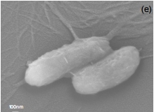

Image of Pseudomonas aeruginosa obtained by scanning electron microscopy: when anionic and cationic polymers are released, they interact with the outside walls of the bacteria to form bundles and cause cell wall disaggregation (International Journal of Molecular Sciences)
Lipid fragments coated with polymers displayed action against three species of bacteria associated with hospital infection.
Lipid fragments coated with polymers displayed action against three species of bacteria associated with hospital infection.

Image of Pseudomonas aeruginosa obtained by scanning electron microscopy: when anionic and cationic polymers are released, they interact with the outside walls of the bacteria to form bundles and cause cell wall disaggregation (International Journal of Molecular Sciences)
By Karina Toledo
Agência FAPESP – Nanoparticles created at the University of São Paulo’s Chemistry Institute (IQ-USP) in Brazil from polymer-coated lipid fragments have displayed potent action against strains of multidrug-resistant bacteria in laboratory experiments.
The results, which were recently published in the International Journal of Molecular Sciences, were presented by Ana Maria Carmona-Ribeiro, the project’s principal investigator, on October 16 during the Brazil-Sweden Workshop on Frontier of Science and Education.
“In previous studies we found both a cationic (positively charged) lipid and a cationic polymer to be active against microorganisms,” Carmon-Ribeiro told Agência FAPESP. “When these two elements are combined or one of them is combined with an antibiotic or an antimicrobial peptide, a broader spectrum of action is obtained.”
Particle sizes ranged from 50 to 100 nanometers (nm). In comparison, a single human hair is approximately 80,000 nm in diameter. The various layers were assembled by electrostatic attraction.
“First we dispersed synthetic cationic lipids to form positively charged bilayer fragments,” she said. “Then we deposited carboxymethylcellulose (CMC), a natural anionic (negatively charged) polymer. Finally, we deposited another layer of a cationic polymer called polydiallyldimethylammonium chloride.”
During master’s and PhD research performed by Letícia Dias de Melo Carrasco under Carmona-Ribeiro’s supervision and with support from FAPESP, the nanoparticles were tested on cultured bacteria that are typically found in hospitals and have developed resistance to the most widely used antibiotics. Some of the bacteria tested included multidrug-resistant Pseudomonas aeruginosa, carbapenemase-producing Klebsiella pneumoniae, and methicillin-resistant Staphylococcus aureus.
According to the results published in the International Journal of Molecular Science, the nanoparticles killed between 92% and 99% of the cultured cells.
“We observed that the nanoparticles disassembled on interacting with the bacteria. The anionic and cationic polymers were released and interacted with the outside walls of the bacteria to form bundles. This caused cell wall disaggregation,” Carmona-Ribeiro said.
Immunologic adjuvants
In previous research, the IQ-USP group combined cationic lipid fragments with pathogen antigens and tested their effectiveness as immunologic adjuvants.
In one experiment, the particles were combined with a protein produced by Mycobacterium leprae, the bacterium that causes leprosy.
“We tested this combination in mice and found that this immunologic adjuvant triggers excellent cellular responses, enhancing the host cell’s capacity to combat the microorganism that is causing the infection,” Carmona-Ribeiro said.
Trials were also performed using ovalbumin, which is the main protein in chicken egg whites and is considered a model antigen for new adjuvant testing. “These assays showed that our adjuvants are better than those currently available in the marketplace,” Carmona-Ribeiro said. "We were limited by the difficulty of finding genuinely good antigens to further test this system.”
A different line of research consisted of combining the cationic lipid fragments with amphotericin B, an antifungal agent used to treat diseases such as aspergillosis, blastomycosis, disseminated candidiasis, coccidioidomycosis, cryptococcosis, histoplasmosis, mucormycosis, disseminated sporotrichosis, and even leishmaniasis.
“We administered this compound parenterally to mice and observed a better effect than that of commercial formulations of amphotericin, such as Fungizone, for example. These fragments are promising, but to continue advancing we need new collaborations with groups who use other in vivo models,” Carmona-Ribeiro said.
Cultivating partnerships
Organized by FAPESP under the aegis of cooperation agreements with Uppsala University and Lund University, the workshop aimed to stimulate new partnerships between researchers in São Paulo State and Sweden by promoting an exchange of research, technologies and experience. The three institutions signed cooperation agreements in 2015.
In addition, in the session entitled “Frontiers in Physical and Biological Sciences,” Watson Loh, a researcher at the University of Campinas’s Chemistry Institute (IQ-UNICAMP), presented his research focused on the creation of synthetic particles that mimic biological systems.
“We combine commercial chemicals like surfactants and water-soluble polymers, in an endeavor to understand how these substances interact to form structures that resemble those found in living things,” Loh said.
One of the future possibilities, he explained, would be to use these particles as drug scaffolds. To this end, the group is working with responsive systems, i.e., systems capable of responding to external stimuli such as temperature, acidity or light.
“The ideal model would be to create a particle capable of going to the exact site at which the drug is to be released and at that moment we would furnish the stimulus,” Loh said. “Factors such as particle size, and both internal and external structure, will determine the particle’s behavior and speed, as well as the site to which it adheres.”
Tommy Nylander, a professor of physical chemistry at Lund University, also presented studies on molecular drug scaffolds, but in this case based on lipids found in cell membranes.
Leif Kirsebom, a professor of biology and head of Uppsala University’s Biomedical Center, presented studies on the genomes of the more than 150 species of bacteria from the genus Mycobacterium. His group is investigating how the expression of important genes varies when these microorganisms are exposed to stresses such as very high or very low temperatures, hypoxia or oxidative stress.
The other participants in the session were Katarina Edwards (Uppsala University), Sylvio Canuto (USP), Sven Lidin (Lund University), Isabel Alves dos Santos (USP), Olov Sterner (Lund University), Eduardo Gonçalves Ciapina (São Paulo State University, Unesp), Jorge Melendez (USP), Andreas Korn (Uppsala University), Jacques Raymond Daniel Lépine (USP), Laerte Sodre Junior (USP), and João Evangelista Steiner (USP).
Republish
The Agency FAPESP licenses news via Creative Commons (CC-BY-NC-ND) so that they can be republished free of charge and in a simple way by other digital or printed vehicles. Agência FAPESP must be credited as the source of the content being republished and the name of the reporter (if any) must be attributed. Using the HMTL button below allows compliance with these rules, detailed in Digital Republishing Policy FAPESP.


