


Graphic summary of the study (image: International Journal of Biological Macromolecules)
Findings published in the International Journal of Biological Macromolecules could guide studies aimed at treating diseases such as osteoporosis.
Findings published in the International Journal of Biological Macromolecules could guide studies aimed at treating diseases such as osteoporosis.
Graphic summary of the study (image: International Journal of Biological Macromolecules)
By Fernanda Bassette | Agência FAPESP – A recent discovery could transform our understanding of bone health maintenance and pave the way for potential treatments for bone diseases, including osteoporosis. Researchers at the University of São Paulo’s Ribeirão Preto School of Dentistry (FORP-USP) in Brazil identified a protein called agrin that plays an essential role in preserving bone mass and quality.
Osteocytes originate from osteoblasts, which are responsible for bone formation, and function as regulators of bone tissue. They help maintain the tissue’s internal balance, a process called homeostasis that is essential for bones to remain strong throughout life.
Until now, it was known that agrin played an important role in regenerating heart muscle and forming cartilage. A group led by Professor Márcio Mateus Beloti from the Department of Basic and Oral Biology at FORP-USP, linked to the Bone Research Lab, was the first to investigate whether this same protein played a similar role in bone tissue.
“What motivated us to investigate the role of agrin in bone is that both bone and heart tissues originate from the same type of cell, called mesenchymal. In other words, during the development of the body, muscles and bones have a common cellular origin and, for this reason, perhaps the agrin protein also plays a role in bone tissue regeneration,” explains Beloti.
In a 2021 study, Beloti’s group demonstrated that osteoblasts produce the agrin protein. Osteoblasts are cells responsible for forming the mineralized matrix of bone tissue. The researchers then conducted an experiment to determine the consequences of eliminating the protein from these cells. They discovered that, without agrin, the osteoblasts differentiated less and consequently formed less mineralized bone matrix.
“This told us the following: osteoblasts with active agrin are physiologically forming bone tissue. If these cells stop producing this protein due to illness or some other reason, there’ll be problems with bone tissue formation,” he explains.
Based on these findings, Professor Beloti’s group investigated a second type of bone cell: osteocytes, which are more mature osteoblasts at the terminal stage of cell differentiation. Researchers Maria Paula Oliveira Gomes, Letícia Faustino Adalpho, and co-supervisor Adalberto Luiz Rosa raised the following hypothesis: Could the agrin protein be produced by osteocytes and play an important role in their function?
To investigate this possibility, the team used genetically modified mice, known as “knockout” mice, in which agrin production was specifically deactivated in osteocytes (conditional knockouts). These were the bone cells of interest. This approach was necessary because the total absence of agrin leads to death at birth due to severe respiratory complications. Therefore, it would be impossible to study the effects of agrin deficiency in bone in an animal completely devoid of the protein.
The group created an animal model in which the agrin protein was removed only from osteocytes. This allowed the researchers to analyze the role of agrin in bone specifically, without affecting its production in other parts of the body. In parallel, the researchers used CRISPR-Cas9 technology to silence agrin in an osteocyte lineage grown in a laboratory. This allowed them to investigate the effects of the protein’s absence in a controlled environment. “We then observed what happened to the bone of this animal,” said Beloti. The study, supported by FAPESP through four projects (2020/14950-4, 2021/03204-2, 2021/04824-4 and 2022/02461-4) was the subject of an article published in the International Journal of Biological Macromolecules.
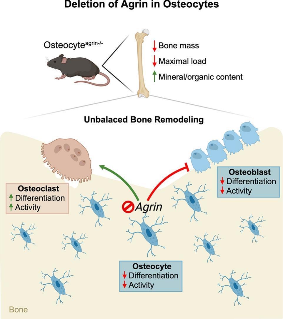
The genetically modified mice were monitored from birth and evaluated again at six weeks of age. Then, they were compared with non-genetically modified animals in the control group. The results were clear: osteocytes produce the agrin protein, and its absence directly impacts bone health.
Less bone tissue
The mice without agrin in their osteocytes showed a significant reduction in bone mass, as well as physical and chemical changes to their bone composition. These changes compromised the tissue’s structure, making the bones more fragile and susceptible to fractures. “Even though they were the same age, the animal without agrin in the osteocytes showed significant differences in bone mass composition compared to the control animal,” said the professor.
In terms of volume, the loss of bone mass was approximately 30%. The bone density (which reflects how compact and resistant the bone is) was also around 30% lower. In a fracture test, which measures the force needed to break a bone, the researchers found that bones without agrin required 15% less force to break.
The researchers then assessed what was happening at the cellular level to cause the lack of agrin to lead to this loss of bone mass and consequent fragility. In the laboratory, the absence of agrin disrupted the maturation and function of osteocytes. This impacted the balance of other bone cells by reducing the activity of osteoblasts, which form bone, and increasing the activity of osteoclasts, which degrade bone.
Therapeutic potential
According to Professor Beloti, the results reinforce the idea that the agrin protein is a fundamental link in the communication circuit between osteocytes, osteoblasts, and osteoclasts, making it a possible target for new therapies. The protein helps preserve tissue integrity by regulating this cellular balance, even in cases where bone loss is not directly related to the absence of agrin. “Stimulating its production in cells could create a mechanism to compensate for bone tissue damage,” the professor suggests.
Despite these advances, it is not yet known if any diseases naturally lead to lower agrin protein production in humans. The next step in the research will be to investigate the possible link between agrin and osteoporosis development. The goal is to determine whether the absence of the protein exacerbates the effects of osteoporosis, a disease that weakens bones and affects millions of people worldwide, particularly the older population.
The article “Agrin-deficient osteocytes disrupt bone tissue homeostasis in male mice” can be read at: www.sciencedirect.com/science/article/abs/pii/S0141813025031034?via%3Dihub.
The Agency FAPESP licenses news via Creative Commons (CC-BY-NC-ND) so that they can be republished free of charge and in a simple way by other digital or printed vehicles. Agência FAPESP must be credited as the source of the content being republished and the name of the reporter (if any) must be attributed. Using the HMTL button below allows compliance with these rules, detailed in Digital Republishing Policy FAPESP.
