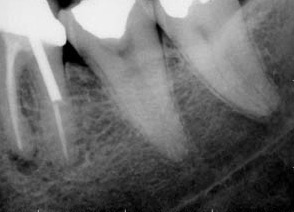

For use in odontology, the laser scanner will allow Brazil to explore new radiology technology that has been mastered by few countries, says project team
For use in odontology, the laser scanner will allow Brazil to explore new radiology technology that has been mastered by few countries, says project team.
For use in odontology, the laser scanner will allow Brazil to explore new radiology technology that has been mastered by few countries, says project team.

For use in odontology, the laser scanner will allow Brazil to explore new radiology technology that has been mastered by few countries, says project team
By Elton Alisson
Agência FAPESP – A group of researchers in São Paulo are developing the first prototype for a digital X-ray machine using Brazilian technology. The project is a partnership between researchers at the Optics and Photonics Research Center (Cepof) at Universidade de São Paulo’s São Carlos Physics Institute (USP – IFSC), Fortaleza’s Instituto Atlântico and Gnatus, a Ribeirão Preto-based industry.
The researchers opted to develop equipment focused on applications in odontology because it is one of the health areas with the highest demand for X-rays. According to researchers participating in the project, the equipment will allow for the development of this new technology, which is transforming the way radiology is performed worldwide, on Brazilian soil.
“Digital radiology is only just beginning. We know that there is enormous demand for this technology in Brazil and that the radiography plates used today for X-ray exams will become obsolete. For this reason, we intend to aid the country’s health system by replacing that technology,” comments Vanderlei Bagnato, professor at IFSC and coordinator at Cepof, an FAPESP Research, Innovation and Dissemination Center (RIDC).
According to Bagnato, the equipment is a laser scanner that performs the same function as those manufactured by few equipment industries in the health field and used by some hospitals in Brazil for digital radiology.
The scanner reads and digitalizes X-ray images obtained from plates composed of rare earth salts, among other materials.
When the X-rays hit these plates, the electronic charges in the molecules that comprise the material are excited and enter a metastable state of energy (which is different from a state of rest). Afterwards, laser irradiation via scanning (similar to that developed by Brazilians) of the X-ray-exposed plates causes the molecules to receive more energy, reaching a state that allows them to return to their previous state and emit a certain quantity of blue light from every point of the film in proportion to the X-ray load.
The scanner reads and submits the image generated by the plate in real time to a high-resolution monitor – much like an ultrasound exam. A specific software processes and generates high-resolution radiography, which can then be stored or sent via the internet.
“Digital radiography almost allows us to perform microscopy using X-rays with images on a practically molecular level,” Bagnato affirmed. “Everything depends on how finely we manage to focus the laser reader.”
National technology
According to the researcher, another advantage this technology offers is reducing the health risks associated with radiation exposure for patients and health professionals. This because the team intends to study ways of using between 50% and 80% fewer X-rays than the conventional method, in addition to eliminating the chemicals used in traditional radiology to develop the photographic film.
Although it is a major technological trend, only few countries in the world – including the United States and Holland – produce the plates and scanner for digital X-rays.
As there are still no manufacturers of the plates or optical scanner in Brazil, according to Bagnato, the Brazilian Development Bank (BNDES) projects a need for the country to invest in digital X-ray technology and has approved the development of the scanner and other parts of the equipment through its Technology Fund (Funtec).
“The digital X-ray must have a source of X-rays, which Brazil knows how to produce and does relatively well,” stated Bagnato. “The scanner would then have to be developed,” he explained.
One of the improvements that researchers intend to make to the existing technology is increasing the sensitivity of the reader through changes in the manner and the geometry of the detection so that the equipment can read films exposed to lower levels of X-rays. This would make it possible to reduce the risks of X-ray exposure to pregnant women and children, who are indicated as the main risk groups for radiation exposure.
“The idea is for the dose of X-rays needed for digital radiology to be so low that it does not create a risk to these people,” explained Bagnato.
The researchers also developed software for digital radiography processing with new applications to allow odontology health professionals to not only visualize radiography but also to obtain such information as bone density and damage to a given tooth.
According to Bagnato, the next step after the conclusion of the prototype phase is to transform the equipment into a marketable model. To this end, the image processing software is already being inserted in the scanner so that a regular microcomputer can read the images.
“If we had to manufacture a microcomputer with the software and laser reader, it would make the equipment very expensive. Our idea is to sell the image processing software that serves as the interface between the laser reader and the microcomputer separately so that the health professional can buy the application and install it in any machine,” he explained.
The researchers are also focused on developing other versions of the equipment for orthopedics and the thorax, for example, over the coming years.
“We are filing an equipment patent focused on odontology that can help to put the country at the forefront of digital radiology development,” stated Bagnato.
Republish
The Agency FAPESP licenses news via Creative Commons (CC-BY-NC-ND) so that they can be republished free of charge and in a simple way by other digital or printed vehicles. Agência FAPESP must be credited as the source of the content being republished and the name of the reporter (if any) must be attributed. Using the HMTL button below allows compliance with these rules, detailed in Digital Republishing Policy FAPESP.


