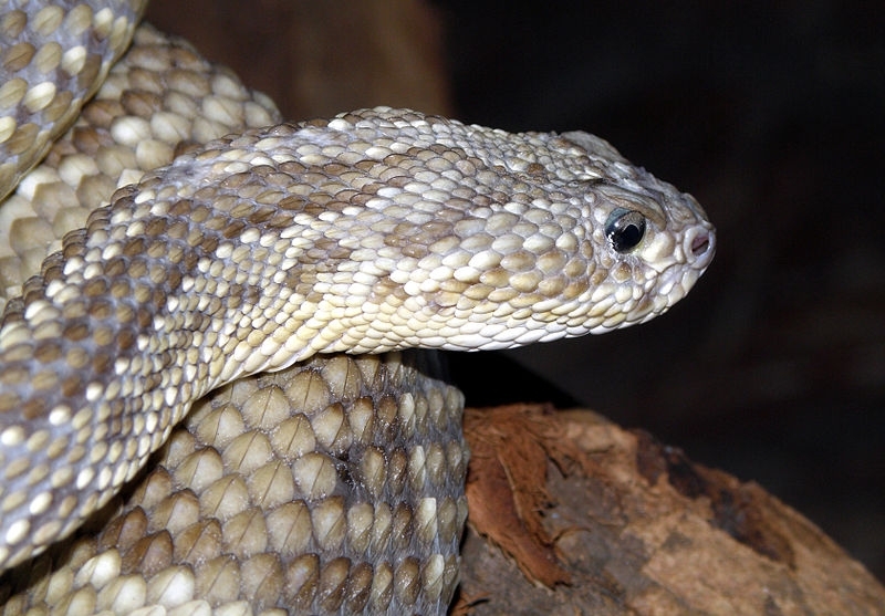

Found in rattlesnake venom, crotoxin can paralyze muscles for a long time and has anti-inflammatory, analgesic, anti-tumoral and immunomodulatory properties (photo: rattlesnake / Wikimedia Commons)
Found in rattlesnake venom, crotoxin can paralyze muscles for a long time and has anti-inflammatory, analgesic, anti-tumoral and immunomodulatory properties.
Found in rattlesnake venom, crotoxin can paralyze muscles for a long time and has anti-inflammatory, analgesic, anti-tumoral and immunomodulatory properties.

Found in rattlesnake venom, crotoxin can paralyze muscles for a long time and has anti-inflammatory, analgesic, anti-tumoral and immunomodulatory properties (photo: rattlesnake / Wikimedia Commons)
By Karina Toledo | Agência FAPESP – Crotoxin is a substance found in the venom of the South American rattlesnake (Crotalus durissus) and is a more potent muscle relaxant than botulinum toxin. In addition, laboratory experiments have shown that crotoxin has anti-inflammatory, analgesic, anti-tumoral and immunomodulatory properties.
However, crotoxin’s therapeutic potential cannot be leveraged for drug development until its interaction with human cells is understood in detail. Important progress in this field of study has been described by Brazilian researchers in an article published in Scientific Reports, an online journal that is owned by Springer Nature.
“We suggest a new structural arrangement of the molecule as well as a model to explain its toxic effect on the nervous system. This information can help other researchers design synthetic compounds with a similar structure and activity to those of the regions of crotoxin that are pharmacologically interesting,” said Carlos Fernandes, a researcher at São Paulo State University’s Botacatu Bioscience Institute (IBB-UNESP) in Brazil.
The study was performed during Fernandes’s postdoctoral research, with FAPESP’s support and supervision by Marcos Roberto de Mattos Fontes, a professor at IBB-UNESP.
“Crotoxin is considered a heterodimer – a complex formed by two different proteins: CA, which is non-toxic and non-enzymatic, and CB, a phospholipase responsible for the neurotoxic effect,” said Fontes, who has been investigating the molecule’s mechanism of action for at least a decade with FAPESP’s support.
In 2008, the group described the three-dimensional structure of the isolated CB protein in the journal Proteins.
Using X-ray diffraction crystallography, a technique that involves crystallizing a substance and observing how the crystal diffracts a beam of incident X-rays, the researchers at IBB-UNESP discovered that when CB is not connected to CA, it tends to cluster in groups of four, forming what scientists call tetramers. In this arrangement, however, the protein’s neurotoxic action is less potent than when it is associated with CA.
A few years later, a group of researchers in France published an article in the Journal of Molecular Biology that described the crystal structure of the complex formed by CA and CB. However, some important regions of CB were hidden by CA in this crystallographic model, including those considered pharmacologically active in the scientific literature: the N-terminal portion (the first amino acid residues in the chain), the C-terminal region (the last residues), and the catalytic site (where the enzymatic reaction occurs).
“This paper refuted our hypothesis that isolated CB organized into tetramers, creating an impasse in the academic community,” Fontes said.
The group at IBB-UNESP then decided to investigate the structural arrangement of the protein complex in solution, i.e., in a liquid medium, a state that more closely resembles the conditions in which the molecule is found in nature.
To perform this examination, they combined five different biophysical techniques: isothermal titration calorimetry, fluorescence spectroscopy, circular dichroism spectroscopy, dynamic light scattering, and small-angle X-ray scattering.
Researchers at the University of São Paulo’s Physics Institute (IF-USP) and Ribeirão Preto School of Philosophy, Science & Letters (FFCLRP-USP), the Federal University of Minas Gerais’s Biological Science Institute (ICB-UFMG) and the Ezequiel Dias Foundation (FUNED) in Belo Horizonte, Minas Gerais, collaborated with the group at IBB-UNESP on this study.
“Our calorimetric experiments showed that the molecules of CB isolated in a recipient formed tetramers. However, as we injected CA into the medium the tetramers dissociated and the two proteins bonded to form heterodimers,” Fernandes said.
This observation was confirmed by dynamic light scattering and small-angle X-ray scattering, both of which measure molecule size. “We measured their diameters and confirmed that isolated CB formed tetramers, CA formed monomers, and the two together formed dimers,” he said.
Fluorescence spectroscopy was then used to discover which parts of the molecule are exposed to the solvent, which in this case was water, Fontes explained. The analysis showed that four residues of the amino acid tryptophan were at the interface between CA and CB. Two of these residues were located at the entrance to the CB catalytic site, blocking access to the region with the protein that is responsible for the biological effect. The N-terminal region of CB was exposed to the solvent, even when the protein bound CA.
“According to the hypothesis we arrived at on the basis of these analyses, two of the toxic regions of CB are practically blocked by CA when they form the heterodimer, but as it approaches the cell membrane, CB is able to bind to the tissue via the N-terminal portion. As a result, CA separates from the complex, enabling the other active regions of CB – the catalytic site and the C-terminal – to bind to the membrane as well, triggering the neurotoxic effect,” Fernandes said.
Presynaptic membrane
Although crotoxin is able to bind to the membrane of any cell in the human body, it predominantly affects the presynaptic membranes located at the junctions between muscles and nerves.
According to Cristiano Oliveira, a researcher at IF-USP and a co-author of the article, the molecule is considered to be one of the main paralyzing agents in rattlesnake venom and has been studied in animal models for the treatment of strabismus because its effects last longer than those of botulinum toxin.
“Previous studies with animals suggested that crotoxin has anti-inflammatory, anti-tumoral and analgesic properties. It may also have esthetic applications,” Oliveira said. “However, it remained to discover how it acts. We describe a possible mechanism of action, which is an important step toward drug development.”
According to Fernandes, the discovery also paves the way for the development of compounds that inhibit the action of crotoxin, which would be useful to enhance the efficiency of snake antivenom.
The article “Biophysical studies suggest a new structural arrangement of crotoxin and provide insights into its toxic mechanism” can be read at: nature.com/articles/srep43885.
Republish
The Agency FAPESP licenses news via Creative Commons (CC-BY-NC-ND) so that they can be republished free of charge and in a simple way by other digital or printed vehicles. Agência FAPESP must be credited as the source of the content being republished and the name of the reporter (if any) must be attributed. Using the HMTL button below allows compliance with these rules, detailed in Digital Republishing Policy FAPESP.


