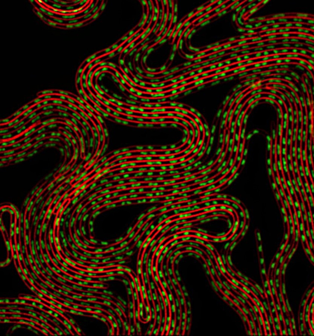

Cells of Bacillus subtilis forced to produce the protein MciZ are unable to divide and transform into long multinucleated filaments. Cell membranes are marked red; the bacterium's DNA is marked green (image: release)
Results published in PNAS can inspire the development of synthetic inhibitors and pave the way for a new class of antibiotics.
Results published in PNAS can inspire the development of synthetic inhibitors and pave the way for a new class of antibiotics.

Cells of Bacillus subtilis forced to produce the protein MciZ are unable to divide and transform into long multinucleated filaments. Cell membranes are marked red; the bacterium's DNA is marked green (image: release)
By Karina Toledo
Agência FAPESP – In an article published on April 6 in the journal Proceedings of the National Academy of Sciences, researchers at the University of São Paulo’s Chemistry Institute (IQ-USP) describe in detail the mechanism by which a protein called MciZ inhibits bacterial cell division.
The research was conducted within the ambit of the project “Smolbnet 2.0: combining genetics and NMR to dissect fundamental protein-protein interactions for complex bacterial division”, coordinated by Frederico José Gueiros Filho and supported by FAPESP.
“The study of proteins that naturally inhibit bacterial cell division not only helps us understand how cells regulate division but also may inspire the development of synthetic inhibitors and pave the way for a new class of antibiotics. The proteins that make up the cell division apparatus are an attractive therapeutic target that isn’t addressed by currently available antibiotics,” Gueiros told Agência FAPESP.
There are some 20 proteins that specialize in controlling bacterial cell division, Gueiros said. The most important is FtsZ (filamentation temperature-sensitive Z). To study the interactions among these proteins, the IQ-USP research group uses as a model the species Bacillus subtilis, which typically lives in soil and does not cause disease in humans.
“This bacterium is an organism that’s very easy to study and serves as an example for others. The division mechanism is similar in all bacteria, and it always depends on FtsZ, so whatever we learn from Bacillus subtilis is applicable to many other species, including pathogens,” Gueiros said.
When a bacterium begins the reproductive cycle, he explained, molecules of FtsZ interweave to form filaments. These filaments assemble into a contractile ring, which marks the site where division will take place.
This Z-ring, as it is known, attracts other division proteins, which alter the outside wall of the cell. At the moment of division, the cell wall stops expanding along its length and begins to grow inward, forming a septum that separates the progenitor cell into two daughter cells.
To ensure that division does not happen at the wrong time or place, proteins inside the cell act as natural inhibitors, preventing formation of a contractile ring if inappropriate. Among the most important of these inhibitors are MciZ (mother cell inhibitor of FtsZ) and MinC, targets of the project coordinated by Gueiros.
“We knew MciZ binds with FtsZ and this somehow prevents formation of a contractile ring, but in order to understand how this happens at the molecular level, we turned to structural biology techniques,” Gueiros said.
These included X-ray crystallography, which researchers used to reveal the 3D structure of the complex formed by the proteins FtsZ and MciZ, and nuclear magnetic resonance (NMR), which they used to study the 3D structure of MciZ in isolation.
The other members of the group were Ana Carolina Zeri, a researcher affiliated with LNBio, and Andrea Dessen, a researcher affiliated with both the Structural Biology Institute (IBS) in Grenoble, France, and LNBio via FAPESP’s São Paulo Excellence Chair (SPEC) program.
Therapeutic target
“Once we’ve detailed the structure of these protein complexes, we can discover which parts of each protein are touching, and from that, we can deduce the mechanism by which one inhibits the other. The site of their interaction is a target for drug development,” Gueiros said.
Several in vitro biochemical experiments were performed to achieve a complete understanding of how the two proteins interact. The scientists observed that the MciZ molecule binds to the end of the filament precisely where there should be another FTsZ molecule, so that the filament stops growing.
“In the absence of MciZ, filaments made up of FtsZ molecules grow until they reach approximately 40 subunits and then form a contractile ring. A small amount of MciZ, in the proportion of approximately 1 to 20, suffices to shorten the length of the filaments by half, so that a contractile ring can’t be formed and cell division is impossible,” Gueiros said.
According to Gueiros, this is a powerful inhibitor mechanism because it is not necessary for MciZ to bind to each of the subunits of FtsZ. “This feature should be taken into account when designing synthetic inhibitors as it means they can be effective at lower concentrations,” he said.
In a previous study, published in the journal PLoS One in 2013, the group investigated mutations in the chain of amino acids that form the protein FtsZ and prevent it from binding to MinC, another cell division inhibitor.
“In this case, the mechanism is different,” Gueiros said. “MinC is unable to bind to the end of the filament, so it binds to the side of the FtsZ molecule, but, even so, it prevents formation of a contractile ring.”
Despite the potential practical implications of their findings, the main motivation of Gueiros’s group is to understand cellular mechanisms at the most fundamental level.
“Good basic research both advances our knowledge of nature and has been the main source of discoveries that fuel innovation and the development of new technologies,” he said.
The article “FtsZ filament capping by MciZ, a developmental regulator of bacterial division” (doi: 10.1073/pnas.1414242112) can be read at www.pnas.org/content/early/2015/04/02/1414242112.abstract" target=.
Republish
The Agency FAPESP licenses news via Creative Commons (CC-BY-NC-ND) so that they can be republished free of charge and in a simple way by other digital or printed vehicles. Agência FAPESP must be credited as the source of the content being republished and the name of the reporter (if any) must be attributed. Using the HMTL button below allows compliance with these rules, detailed in Digital Republishing Policy FAPESP.


