


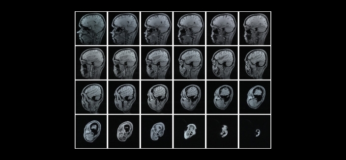
Peptide fragments marked with a radioactive isotope are evaluated as biomarkers of the degenerative process that leads to neuron death (image: Wikimedia Commons)
Peptide fragments marked with a radioactive isotope are evaluated as biomarkers of the degenerative process that leads to neuron death.
Peptide fragments marked with a radioactive isotope are evaluated as biomarkers of the degenerative process that leads to neuron death.
Peptide fragments marked with a radioactive isotope are evaluated as biomarkers of the degenerative process that leads to neuron death (image: Wikimedia Commons)
By Karina Toledo, in Foz do Iguaçu (Paraná) | Agência FAPESP – Research performed at the Medical School of Santa Casa Hospital (FCM-SCSP) in São Paulo, Brazil, with support from FAPESP, could lead to the early diagnosis of Alzheimer’s disease.
At present, no biological markers or scans are available to detect progressive degenerative diseases of the brain in routine clinical practice before there are signs of cognitive decline, and diagnosis is achieved simply by excluding other conditions that cause memory loss and dementia.
“It’s estimated that when patients start to manifest symptoms of cognitive impairment roughly 50% of their neurons have already died. By then, there isn’t much you can do. However, if we could detect the degenerative process when it begins, the chances of stabilizing its progression with the drugs available today would be far greater,” said Luciana Malavolta Quaglio, a professor in FCM-SCSP’s Department of Physiological Sciences.
Some of the results of the research led by Malavolta were presented on August 30 during the 31st Annual Meeting of the Federation of Experimental Biology Societies (FeSBE), held in Foz do Iguaçu, Paraná State.
In her laboratory, Malavolta synthesized small peptide fragments capable of being attracted by beta-amyloid, a larger peptide that plays a crucial role in the development of Alzheimer’s.
For reasons scientists do not yet fully understand, in individuals with this disease, the beta-amyloid molecules naturally present in the organism cluster together, forming beta-amyloid plaques. These plaques build up in the brain and cause a series of alterations that, in conjunction with other factors, result in the death of neurons.
The aim of Malavolta’s research is to develop biomarkers that can signal the presence of beta-amyloid plaques in the brain during a clinical examination.
“We’re testing four different peptide fragments, all of which have few amino acids,” Malavolta told Agência FAPESP. “Whereas the beta-amyloid peptide contains some 42 amino acid residues, ours have between four and six. If they were large, they wouldn’t reach the brain because they wouldn’t be able to cross the blood-brain barrier, a mass of tightly packed cells that protect the central nervous system from potentially toxic substances in the blood.”
The process of designing the molecules was completed in 2011. Since then, in collaboration with scientists at the Albert Einstein Jewish Education & Research Institute (IIEP) in São Paulo, Malavolta has been developing enhanced methods of radiomarking in which the peptide fragments bind with radioactive isotopes so that the distribution of the compound in the organism can be monitored and scans can be performed.
The strategy is similar to the use of scintigraphic scans to assess kidney or heart function. A radiomarked compound with affinity for the tissue of interest is injected into the organism. When the elements reach the target organ, emitted radiation is identified by a scintillation chamber, which converts it into images that specialists can interpret.
Radiomarking is traditionally performed with technetium-99m, a metastable isotope of the element technetium that emits gamma rays. According to Malavolta, this isotope is widely used in nuclear medicine for diagnostic imaging purposes because it has a half-life of six hours, enough to perform a scan and for the patient to be discharged from the hospital on the same day.
“According to the scientific literature, radiomarking techniques using direct labeling of a molecule with technetium achieve yields of between 60% and 65%, in terms of the percentage of fragments that remain bonded to the radioisotope. We have obtained values in excess of 90%, which is considered highly satisfactory in the field of nuclear medicine,” she said.
Preclinical trials
Several in vitro and in vivo trials were performed to evaluate the stability of the radiomarked peptides and their biodistribution in the organism.
One experiment compared a group of healthy mice with a group that had been genetically modified to develop a condition similar to Alzheimer’s. In this model, beta-amyloid plaque formation is induced in the animal’s brain by inserting into its genome a double mutation in the gene encoding amyloid precursor protein (APP), which gives rise to beta-amyloid peptide.
The radiomarked fragments were injected into both groups of mice, and after different periods of time, the researchers measured the radiation in each organ using a gamma-ray counter.
“Depending on the fragment, we observed that between 3% and 5% of the radiomarked molecules reached the brains of the GM mice,” Malavolta said. “This proportion is considered satisfactory. Radiopharmaceuticals currently used in other kinds of diagnosis have a specificity of about 1%.”
In the healthy animals, which served as a control group, only 0.5% of the radiomarked peptides reached the brain.
The in vitro study gave a 50% rate of interaction between radiomarked fragments and brain cells from mice with Alzheimer’s, compared with 10%-12% for the control group.
The rate of binding between radiomarked fragments and murine plasma proteins was about 35% for both groups.
“In this case, the lower the rate, the better, because a larger proportion of the compound is left free to reach the desired target,” Malavolta said. “The result of the experiment shows that 65% of our peptide fragment remained free to travel throughout the organism. The plasma protein-binding rate for some of the drugs currently available is 95%. This means only 5% of the molecules are free, yet even so they’re effective to some extent. If 65% of the compound is free, it’s obviously going to be much more effective.”
One of the strategies Malavolta plans to test to increase the percentage of radiomarked fragments that reach the brain is encapsulation in nanoparticles. Initial testing has already begun.
Preliminary results of the research presented at FeSBE have also been published in Neurological Sciences, Neuropeptides, Journal of Peptide Science, Protein & Peptide Letters and Revista Brasileira de Psiquiatria, among others.
The Agency FAPESP licenses news via Creative Commons (CC-BY-NC-ND) so that they can be republished free of charge and in a simple way by other digital or printed vehicles. Agência FAPESP must be credited as the source of the content being republished and the name of the reporter (if any) must be attributed. Using the HMTL button below allows compliance with these rules, detailed in Digital Republishing Policy FAPESP.
