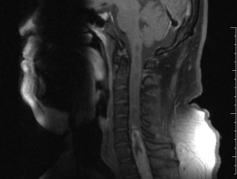

Rare tumor found in 33-year-old patient's spinal cord at Barretos Cancer Hospital in São Paulo State, Brazil (image: PLoS One)
Group identifies chromosomal alterations and genetic mutations associated with rosette-forming glioneuronal tumor of the fourth ventricle.
Group identifies chromosomal alterations and genetic mutations associated with rosette-forming glioneuronal tumor of the fourth ventricle.

Rare tumor found in 33-year-old patient's spinal cord at Barretos Cancer Hospital in São Paulo State, Brazil (image: PLoS One)
By Karina Toledo
Agência FAPESP – Researchers at the Barretos Cancer Hospital in São Paulo State, Brazil, have produced a detailed genetic profile of a rare type of brain cancer called a rosette-forming glioneuronal tumor of the fourth ventricle. Their findings were recently published in the journal PLoS One.
According to Rui Manuel Reis, principal investigator for the study, which was supported by FAPESP, fewer than 100 cases of this cancer have been reported in the literature. This was the first scientific report of a case occurring in Brazil.
“Based on previously published reports, we knew that it’s an indolent tumor with a good prognosis that is found in young adults,” Reis said. “Only some immunohistochemical markers have been identified. That’s it. In our study, we set out to characterize this tumor genetically, locate its origin, and understand the chromosomal alterations and mutations involved.”
The study was performed during Lucas Tadeu Bidinotto’s postdoctoral research, supervised by Reis and also supported by FAPESP.
Reis said rosette-forming glioneuronal tumors of the fourth ventricle were not classified as pathological entities by the World Health Organization (WHO) until 2007. Before that, every case reported used a different nomenclature. The name, now considered official, refers to the mixed nature of the tumor, which involves both neurons and glial cells, and alludes to the structures formed by the tumor cells, which resemble the petals of a flower.
Whereas in most reported cases this type of tumor has been found in the fourth brain ventricle, a cavity located in the brain stem just in front of the cerebellum, the 33-year-old patient treated at Barretos Hospital presented with a tumor located in the spinal cord. Only two other events like this have been described previously.
A sample was taken from the tumor and analyzed using a variety of methods. The group used a technique called microarray-based comparative genomic hybridization (array CGH)to study all tumor cell chromosomes in search of genetic alterations.
“There are significant chromosomic alterations in this tumor in regions that aren’t typical of brain tumors,” Reis said. “For example, there are gains in chromosomes 9 and 16. In other words, instead of two copies we found four. We also identified losses in the number of copies of chromosome 1.”
When the researchers analyzed the DNA present in blood samples from the same patient and compared it with the tumor cells, they found the alterations only in the latter, indicating that the changes had occurred after birth.
“Although most of the alterations we found are rare in brain tumors, the one observed in chromosome 7 was very familiar. It was a fusion of two genes, BRAF and KIAA1549,” Reis said. This finding was confirmed by three other techniques: reverse transcription polymerase chain reaction (RT-PCR), fluorescence in situ hybridization (FISH), and DNA sequencing.
In a previous study published in the Journal of Neuropathology and Experimental Neurology, the group found this fusion in 60% of patients with pilocytic astrocytoma, a brain tumor that originates from star-shaped brain cells called astrocytes and occurs mainly in children and young adults. Its presence is associated with a good prognosis (read more at http://agencia.fapesp.br/21918).
“This finding allows us to raise the hypothesis that rosette-forming glioneuronal tumors of the fourth ventricle have some etiological relationship with pilocytic astrocytomas, which are also indolent tumors that affect the young,” Reis said.
Mutated genes
Next-generation sequencing was used to profile the mutations present in the tumor with an initial panel of 20 target genes identified in the context of a glial cell research project funded by FAPESP. This was followed by sequencing of the exome, i.e., all protein-coding genes in the genome (approximately 2% of the total).
“None of the 20 target genes had mutated, so we decided on exome sequencing and found four somatic mutations, which were later confirmed by the traditional sequencing method,” Reis said. Somatic mutations are acquired after conception and are present only in non-germline cells.
One of the mutated genes was SCN1A, which is associated with hereditary forms of epilepsy. Another was MLL2, which has recently been linked to the development of medulloblastoma and appears to regulate the way in which genes are expressed through epigenetic mechanisms, such as DNA methylation.
The researchers also found mutations in CNNM3 and PCDHGC4 that had never been described before in the literature.
“The next step should be to validate these results in other tumors of the same type in order to try to establish whether these four genes are involved in the development of rosette-forming glioneuronal tumors. We plan to get in touch with researchers who have reported this type of tumor to see if we can design a study in collaboration with them,” Reis said.
The article “Molecular Profiling of a Rare Rosette-Forming Glioneuronal Tumor Arising in the Spinal Cord” (doi: 10.1371/journal.pone.0137690) can be read at journals.plos.org/plosone/article?id=10.1371/journal.pone.0137690.
Republish
The Agency FAPESP licenses news via Creative Commons (CC-BY-NC-ND) so that they can be republished free of charge and in a simple way by other digital or printed vehicles. Agência FAPESP must be credited as the source of the content being republished and the name of the reporter (if any) must be attributed. Using the HMTL button below allows compliance with these rules, detailed in Digital Republishing Policy FAPESP.


