


The findings highlight how hunger and food insecurity can affect future generations (image: Freepik)
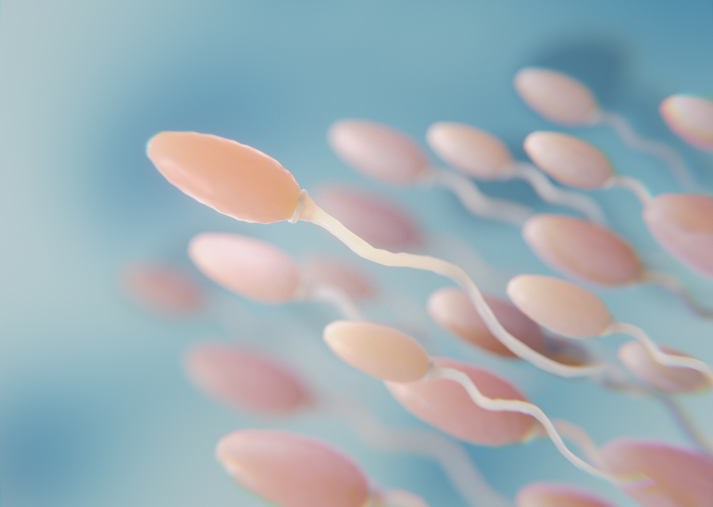
Research with rats has shown that a maternal diet lacking protein during these stages leads to changes in the structure and function of the epididymis, which would explain impairments in motility, viability, and sperm concentration in male offspring.
Research with rats has shown that a maternal diet lacking protein during these stages leads to changes in the structure and function of the epididymis, which would explain impairments in motility, viability, and sperm concentration in male offspring.
The findings highlight how hunger and food insecurity can affect future generations (image: Freepik)
By Maria Fernanda Ziegler | Agência FAPESP – In Brazil, researchers at São Paulo State University (UNESP) conducted experiments on rats and identified that a protein-deficient diet during pregnancy and breastfeeding can compromise the reproductive health of male offspring. A study published in the journal Biology Open found that maternal protein restriction causes changes in the structure and function of the epididymis, an organ in the reproductive system responsible for the maturation and storage of sperm.
Following the offspring into adulthood, the researchers observed that changes in the epididymis were associated with impaired sperm quality, which can compromise fertility. These studies, which were supported by FAPESP, were the first to link maternal protein restriction during pregnancy and lactation to changes in the structure and function of the organ. This helps identify a possible cause of the decline in sperm quality observed in children whose mothers were on this diet (read more at agencia.fapesp.br/35771 and agencia.fapesp.br/50692).
“More and more studies have shown that combating hunger and food insecurity today is also a way to prevent diseases and promote the well-being of future generations. Our work is an example of this. In addition to the diseases associated with the impacts of maternal protein restriction during pregnancy and lactation in rats, we were able to demonstrate that there’s also an impact on the reproductive health of offspring,” says Raquel Fantin Domeniconi, a professor at the Institute of Biosciences (IB-UNESP) in Botucatu.
Studies linking the health of pregnant women to the development of their children have been conducted in recent decades, particularly in a field of research called “developmental origins of health and disease” (DOHaD). There is strong evidence that inappropriate gene-environment interactions during the embryonic phase and the first two years of life are an important factor in the increased incidence of chronic, noncommunicable diseases throughout life, such as cancer, diabetes, chronic respiratory diseases, and cardiovascular diseases.
These epigenetic phenomena have been studied in greater depth using animal models. “An analogy I like to use to explain this problem is that when there’s maternal malnutrition during the embryonic phase, it’s as if there were missing ‘bricks’ in the construction of these organs. This can reduce, for example, the number of nephrons in the kidneys, cells in the heart, and also affect the epididymis, as we saw in our study,” Domeniconi explained to Agência FAPESP.
In the study, pregnant rats were randomly divided into three groups. One group received a normal diet, one group received a diet with 17% protein, and one group received a low-protein diet (6%) during pregnancy and lactation. The researchers found several changes in the epididymis of rodents whose mothers underwent protein restriction. The epididymis is a single duct located behind each testicle. It stores and transports sperm and is where sperm cells mature. It is a hormone-dependent organ that performs specific functions contributing to the creation of an intraluminal environment suitable for concentrating and maturing sperm produced by the testes.
“We found that maternal protein restriction causes significant changes in the epididymis. In addition to reducing the length of the duct, protein deficiency altered its structure and function, affecting the dynamics of luminal fluids, the formation of new blood vessels, and the expression of various proteins,” reports the researcher.
Changes were also observed in the epithelium, the tissue lining the inside of the epididymis, which affects the process of cell differentiation and consequently its function.
The effects of the changes that occurred during the embryonic phase persisted throughout the animals’ lives. Between seven and 14 days of life, while still in the lactation period, a delay in epididymis lining cell differentiation was observed in the offspring of mothers on a low-protein diet. Analysis of these animals’ epididymides showed that they had more mesenchymal cells, which have differentiation potential, than epithelial cells.
These changes all compromise sperm maturation and quality, impacting rats at 44 days of life, which is when the epididymis epithelium is expected to be fully differentiated and the rodents are expected to enter puberty.
“The epididymis is a long, single duct through which sperm pass and mature progressively. When this duct is shortened, the interactions of sperm with the epididymal epithelium and the intraluminal environment are limited, preventing the sperm from becoming functional and capable of fertilizing an oocyte. It’s not that the rodents became completely sterile, but the quality of their sperm is inferior, which could impact fertility,” says the researcher.
For Domeniconi, this finding highlights how hunger and food insecurity can affect future generations. The study showed that a mother’s diet during pregnancy and breastfeeding can have a lasting impact on her children’s genital development. This finding opens the door to further investigation into how nutrition during this period influences reproductive health.
She explains that although it is a structural problem – there is a lack of essential nutrients needed to form the epididymis – most of the changes observed are the result of adaptations of the epididymis to ensure sperm maturation even in a dysfunctional context.
“They’re adaptations upon adaptations so that, even with this new structure, it’s possible for the sperm to mature and maintain their ability to fertilize. After all, from an evolutionary point of view, reproduction is essential for the survival of the species. But if even reproductive capacity is compromised in these cases, it’s worth considering how malnutrition at critical stages can cause profound changes in the body,” Domeniconi points out.
The article “Maternal protein restriction affects the differentiation of cells in the epididymal epithelium lining of 44-day-old rats” can be read at journals.biologists.com/bio/article/14/6/bio060080/368206/.
The Agency FAPESP licenses news via Creative Commons (CC-BY-NC-ND) so that they can be republished free of charge and in a simple way by other digital or printed vehicles. Agência FAPESP must be credited as the source of the content being republished and the name of the reporter (if any) must be attributed. Using the HMTL button below allows compliance with these rules, detailed in Digital Republishing Policy FAPESP.
