


Aerobic exercise regulates the functioning of cardiac cells, restores mitochondrial mechanisms, and improves the heart’s ability to pump blood
Aerobic exercise regulates the functioning of cardiac cells, restores mitochondrial mechanisms, and improves the heart’s ability to pump blood.
Aerobic exercise regulates the functioning of cardiac cells, restores mitochondrial mechanisms, and improves the heart’s ability to pump blood.
Aerobic exercise regulates the functioning of cardiac cells, restores mitochondrial mechanisms, and improves the heart’s ability to pump blood
By Karina Toledo
Agência FAPESP – A recent study conducted by the Universidade de São Paulo (USP) and published in the PLoS One journal reveals that aerobic physical exercise can interrupt the degenerative process observed in cardiac failure, a disease characterized by the heart’s inability to adequately pump blood.
The study is a continuation of previous research in which a group coordinated by Julio Cesar Batista Ferreira, of the Department of Anatomy at USP’s Institute of Biomedical Sciences, discovered that failing hearts do not beat because of a defect in the quality control system of cardiac cells responsible for destroying damaged proteins.
Without this cleaning mechanism, highly reactive proteins accumulate in the cytoplasm, interact with other structures, and eventually cause cardiac cell death, worsening the prognosis.
In a previous study, these scientists tested the βIIV5-3 molecule in rats and found that it was capable of normalizing quality control and reversing the degenerative process.
In the most recent experiment, also conducted on rats and funded by FAPESP, aerobic exercise proved as efficient as pharmacological treatment in reactivation of the “cellular cleaning system”.
“We used the same animal model, which consists of tying one of the rat’s coronary arteries to induce myocardial infarction. The lack of blood irrigation causes immediate death to approximately 30% of cardiac cells. After a month, the animal presents signs of [cardiac] failure,” explains Ferreira.
Although infarction is the main cause of cardiac insufficiency, explains the researcher, this degenerative process is a common final result of different untreated chronic diseases, including hypertension, diabetes, and obesity. This malady leads to the death of approximately 70% of affected patients in the first five years.
“After the primary damage, the remaining cells must work twice as hard to offset the chronic injury. Because they are not prepared for this, they end up collapsing over time,” he said.
To evaluate the impact of aerobic physical exercise on a heart under these conditions, the research team conducted an experiment with 27 rats split into three groups. In the first group, comprising 10 animals, the researchers induced a heart attack and the animals remained sedentary. In the second, the infarction was induced in 8 rodents, which were later submitted to a running program on treadmills for one hour per day, five times per week for six months. The third group, the control group, was subjected to neither induced heart attacks nor exercise.
A series of measurements were undertaken in the third week after the heart attack to confirm heart failure in the rodents. The tests were repeated after eight weeks of physical training.
In the group of “running” rats, heart function – which is the cardiac capacity to pump blood to arteries – improved by 70% when compared to the animals that had suffered heart attacks and remained sedentary. Their exercise tolerance, which is the distance that the animals managed to run on the treadmills, increased from 280 to 760 meters. The maximum consumption of oxygen, which had previously been reduced by 20%, normalized, reaching values equivalent to those of the control group.
To understand how physical exercise benefits the heart, scientists conducted molecular analysis on the organ after the experimental stages. “We wanted to determine whether exercise also modulates the quality control system for protein, and we discovered that it does. As much as drug treatment,” explains Ferreira.
According to the researcher, an intracellular complex known as proteasome is largely responsible for degradation of the damaged proteins. In the previous study, the group found that the heart failure patients typically see activity reduced by more than 50%.
“Physical exercise normalized proteasome activity in the hearts of rats and thereby restored quality control,” explains Ferreira.
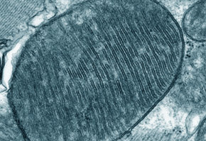
The next step was to discover why proteasome activity was reduced and how it returned to normal with physical training. “This complex requires much energy to work. Because the main producer of energy in cells are the mitochondria, we hypothesized that the mitochondrial metabolism could be compromised,” explained the researcher.
“Molecular analysis conducted on the hearts of animals and cardiac cell cultures showed that, in fact, the mitochondria function well during heart failure.
“The mitochondria begin to consume less oxygen, produce less ATP (adenosine triphosphate, a molecule that stores energy), and generate excess quantities of reactive oxygen and nitrogen species,” comments Ferreira.
These free radicals can react with different molecules in the cell and cause a collapse, affirms the researcher. The oxidation of phospholipids found in the mitochondrial membrane, for example, generates an extremely reactive aldehyde, 4-hydroxynonenal (4-HNE), which directly attacks proteasome and inhibits cardiac failure.
“Physical exercise was capable of restoring the metabolism of mitochondria and, consequently, reducing the release of free radicals and their subproducts in the hearts of animals with cardiac insufficiency,” explains the researcher.
In the hearts of animals that performed physical exercise, 4-HNE levels were lower; consequently, the proteasome was less susceptible to 4-HNE attacks and worked better. “Understanding the integration of intracellular systems is the key to improving our biological understanding of cardiac cells,” said Ferreira.
The experiments were conducted during the master’s studies of Julian Cruz Campos, under the orientation of Ferreira and with a FAPESP fellowship. The study also boasted the participation of researchers Alicia Kowaltowski and Patricia Chakur Brum of USP along with Daria Mochly-Rosen of Stanford University.
Further study
The βIIV5-3 molecule developed by Ferreira’s group in partnership with Stanford deactivates a protein known as PKCβII, which also bonds with the proteasome and affects protein quality control.
Studies to transform βIIV5-3 into a medication continue and, while awaiting funding to begin clinical trials on humans, the researchers are investigating the functioning of molecules in the organism.
In the most recent studies, the researchers found that PKCβI is also one of the factors responsible for the poor function of mitochondria and that βIIV5-3 can help in this case.
“We are attempting to develop a drug that can work on key points of the biology of cardiac cells. βIIV5-3, for example, improves the quality control of proteins and restores the cellular metabolism. However, restoring quality control is useless if there is little ATP in the cell, as the proteasome needs a large amount of energy to function,” said the researcher.
For Ferreira, physical exercise is an important complementary therapy. It can help to reduce medicine dosages and consequently adverse side effects in addition to improving quality of life.
The researcher stressed, however, the importance of being under a doctor’s care for those with cardiac disease who are beginning physical exercise. “Excessive and poorly planned exertion can worsen things instead of helping,” he warns.
The article Exercise Training Restores Cardiac Protein Quality Control in Heart Failure (doi: 10.1371/journal.pone.0052764) can be read in www.plosone.org/article/info%3Adoi%2F10.1371%2Fjournal.pone.0052764.
The Agency FAPESP licenses news via Creative Commons (CC-BY-NC-ND) so that they can be republished free of charge and in a simple way by other digital or printed vehicles. Agência FAPESP must be credited as the source of the content being republished and the name of the reporter (if any) must be attributed. Using the HMTL button below allows compliance with these rules, detailed in Digital Republishing Policy FAPESP.
