


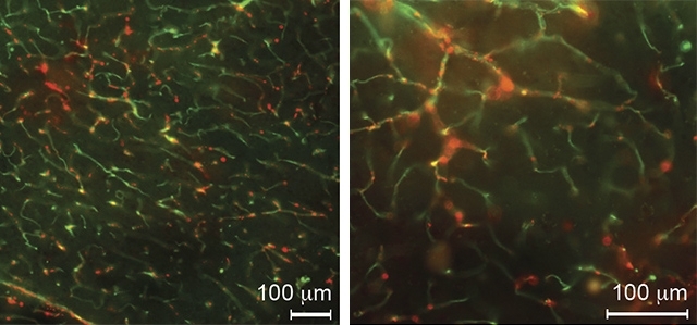
Peptide developed in Brazil is capable of binding to the blood-brain barrier that protects the central nervous system and could help create novel imaging tests to diagnose Alzheimer’s and Parkinson’s (image: phage bound to blood vessels in brain hemispheres of mice is visualized with antibacteriophage sera [red] / Proceedings of the National Academy of Sciences)
Peptide developed in Brazil is capable of binding to the blood-brain barrier that protects the central nervous system and could help create novel imaging tests to diagnose Alzheimer’s and Parkinson’s.
Peptide developed in Brazil is capable of binding to the blood-brain barrier that protects the central nervous system and could help create novel imaging tests to diagnose Alzheimer’s and Parkinson’s.
Peptide developed in Brazil is capable of binding to the blood-brain barrier that protects the central nervous system and could help create novel imaging tests to diagnose Alzheimer’s and Parkinson’s (image: phage bound to blood vessels in brain hemispheres of mice is visualized with antibacteriophage sera [red] / Proceedings of the National Academy of Sciences)
By André Julião | Agência FAPESP – Brazilian and US researchers have developed a molecule called FRW that, in tests with mice, proved capable of binding to blood vessels in the brain but not in other organs when injected into the bloodstream. They used the technique to produce the first ever complete map of the vasculature of the brain, paving the way for the creation of novel diagnostic imaging strategies and therapies for diseases such as Alzheimer’s and Parkinson’s.
The research was supported by FAPESP and led by Ricardo José Giordano, a professor at the University of São Paulo’s Chemistry Institute (IQ-USP) in Brazil and head of its Vascular Biology Laboratory. The results are published in Proceedings of the National Academy of Sciences (PNAS).
As Giordano explained, the main obstacle to the development of drugs capable of binding to blood vessels in the brain is the blood-brain barrier, a selectively permeable cellular boundary that protects the central nervous system from potentially toxic substances in the bloodstream. However, this research shows that FRW binds precisely to endothelial cell junctions in the blood-brain barrier.
In addition to producing a complete map of the brain’s vasculature, therefore, the new technique could also be used to detect the kind of gap in the blood-brain barrier that may be one of the causes of neurodegenerative diseases such as Alzheimer’s and Parkinson’s.
“Theoretically speaking, if FRW doesn’t bind to the cerebrovascular system, it’s a sign that the barrier is impaired,” Giordano told Agência FAPESP.
To perform the study, the researchers used a library of bacteriophages, usually called phages – viruses that infect bacteria and can be used to deliver molecules because they are inoffensive to other organisms.
“Each of the phages in the library has been modified by genetic engineering to have a different surface peptide [protein piece] from that of the original virus. This peptide carries a marker that’s detected when it binds to specific proteins, be they in the cerebrovascular system, in tumors, kidneys or other regions of the organism,” Giordano said.
The technique is known as phage display and won the 2018 Nobel Prize in Chemistry for its creators, George P. Smith and Gregory P. Winter. Smith first described phage display in 1985. In the following decade, the technique was adapted for application to living animals by Renata Pasqualini, a Brazilian-born researcher at Rutgers University in the US and one of the authors of the article in PNAS.
The research led by Giordano began in 2011 as part of a project for which FAPESP awarded a scientific initiation scholarship to Fenny Hui Fen Tang, first author of the article. Tang continued to focus on this research interest for her master’s and again for her doctorate, recently completed at IQ-USP.
FAPESP also provided funding to Giordano via a Young Investigator Grant and a Regular Research Grant.
To arrive at the molecule, the researchers injected mice with an entire library comprising some 10 billion different phages. The modified viruses circulated in the bloodstream, and although most were eliminated by the organism, some bound to the vasculature of different organs and tissues, including the blood-brain barrier.
These phages were retrieved from the animals’ brains and cultured in bacteria so that they multiplied. The next-generation phages were injected into other mice to enhance selection. After three cycles, approximately 3,000 phages bound to blood vessels in the brain.
“The selection process was won by the peptides with the most affinity with the cerebrovascular system because they multiplied the most,” Giordano explained.
Out of the 3,000-odd peptides that bound to the blood-brain barrier, 1,021 contained a sequence of three amino acids: phenylalanine, arginine and tryptophan (FRW).
“We found this sequence to be a panvascular marker for the brain, since it recognizes all brain blood vessels,” Giordano said. “However, it doesn’t bind to blood vessels in other tissues that are also protected by a barrier, such as those in the colon and testes.”
Giordano and his group were surprised to find that FRW did not bind to blood vessels in the retina, hitherto considered an extension of the central nervous system.
“The barrier that protects blood vessels in the retina was believed to be very similar or even identical to the blood-brain barrier, but we discovered a difference, at least in mice, thanks to this molecule,” he said. This finding alone will lead to more research on the blood-retina barrier.
Synthetic molecule
The researchers at IQ-USP tried unsuccessfully to use biochemical techniques to identify the cellular receptor to which the phages bound, so they collaborated with colleagues at Adolpho Lutz Institute in São Paulo who specialize in transmission electron microscopy (TEM). This team not only helped them to see the molecule in the brains of live animals but also demonstrated that the phages bound to endothelial cell junctions in the blood-brain barrier. The junctions concerned are known as “tight” because they prevent all foreign substances, including water, from crossing the blood-brain barrier.
“We now need to study the phenomenon in more detail, as the structure is made up of several molecules,” Giordano said.
The next step will be to synthesize the peptide and see if the version produced in the laboratory acts the same way as FRW in mice. The researchers think the synthetic version also binds to blood vessels in the brain but have not been able to see this in vivo.
Another future direction will be to explore other peptides that do not contain FRW and select those that remain in specific brain regions, such as the cerebellum, olfactory bulb and hemispheres, permitting more specific diagnostic tests in the future.
The article “A ligand motif enables differential vascular targeting of endothelial junctions between brain and retina” (doi: 10.1073/pnas.1809483116) by Fenny H.F. Tang, Fernanda I. Staquicini, André A.R. Teixeira, Jussara S. Michaloski, Gislene M. Namiyama, Noemi N. Taniwaki, João C. Setubal, Aline M. da Silva, Richard L. Sidman, Renata Pasqualini, Wadih Arap and Ricardo J. Giordano can be retrieved from www.pnas.org/content/116/6/2300.
The Agency FAPESP licenses news via Creative Commons (CC-BY-NC-ND) so that they can be republished free of charge and in a simple way by other digital or printed vehicles. Agência FAPESP must be credited as the source of the content being republished and the name of the reporter (if any) must be attributed. Using the HMTL button below allows compliance with these rules, detailed in Digital Republishing Policy FAPESP.
