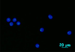

A faster, more efficient method developed by researchers at the NIH and the CTC makes treatment selection easier, especially for leukemia patients. The article was published in Blood
A faster, more efficient method developed by researchers at the NIH and the CTC makes treatment selection easier, especially for leukemia patients. The article was published in Blood.
A faster, more efficient method developed by researchers at the NIH and the CTC makes treatment selection easier, especially for leukemia patients. The article was published in Blood.

A faster, more efficient method developed by researchers at the NIH and the CTC makes treatment selection easier, especially for leukemia patients. The article was published in Blood
By Fábio de Castro
Agência FAPESP – A group of scientists from Brazil and the United States developed a new method to diagnose chromosomal alterations in cancer patients—especially those with leukemia—at a much faster and in a more accurate manner than with conventional cytogenetic technology.
The study, which was published in Blood, involved researchers from the National Institutes of Health (U.S.) and the Cellular Therapy Center (CTC) of Riberão Preto. Coordinated by Marco Antônio Zago, the CTC is one of the FAPESP Research, Innovation and Dissemination Centers (CEPID).
According to the article, the detection of chromosomal anomalies makes it possible to predict potential responses to therapy and is therefore considered one of the primary tools to direct clinical treatment.
“Detecting chromosomal alterations as quickly and precisely as possible is necessary for us to be able to diagnose the cancer and choose the best treatment for the patient,” said researcher Rodrigo Calado, one of the article’s authors and a professor in the Clinical Medicine Department of the Riberão Preto Medical School (FMRP-USP), part of Universidade de São Paulo.
Calado said the conventional cytogenetic method used to detect chromosomal alterations takes a few days to give results and only analyzes approximately 20 tumor cells. The limited number of cells analyzed increases the likelihood of a false negative. “With the method we developed, we can analyze 20,000-30,000 tumor cells in one or two days. We can find chromosomal alterations in a much more precise manner this way,” he said.
With the conventional cytogenetic technique, scientists must examine cells one by one under a microscope, but with the new method, 30,000 cells can be analyzed quickly by flow cytometry—a technology used in many laboratories for other analyses.
“In flow cytometry, hundreds of cells pass through a laser beam every second, which identifies the fluorophores used to analyze the chromosomes. That way, the irregularities are quickly detected,” he stated.
Calado, who returned to Brazil in 2011 after an 8-year period as researcher at the NIH, had already used this method in North American laboratories to evaluate telomeres, which are the extremities of chromosomes.
“We had many difficulties with the limitations of cytogenetic methods because we could only analyze a small number of cells, and especially in cases like myelodysplasia—a type of leukemia—the results were very imprecise, and many chromosomal alterations went unnoticed. So we had the idea of using the telomere evaluation method to analyze the quantitative alterations of the chromosome as a whole,” he explained.
To perform this type of chromosomal analysis, the scientists developed a new method for preparing the cells that is more sophisticated than the conventional preparation method, and they improved the data analysis software.
“The result was an easily applied method because we used a flow cytometer, technology that already exists and is found in many laboratories. Because of this, the new method can be quickly adopted for routine use,” he said.
In its original application, the flow cytometer was used to analyze telomeres, important markers for cell aging, which is a process that is related to a series of diseases.
“This equipment is used in a method that identifies very short telomeres, making it possible to detect diseases such as pulmonary fibrosis and aplastic anemia. Telomere shortening is the best biomarker for aging,” said Calado.
The article Chromosome Flow-FISH by Neal Young, Rodrigo Calado and others may be read by Blood subscribers at: http://bloodjournal.hematologylibrary.org/content/early/2012/08/29/blood-2012-05-434266.abstract.
Republish
The Agency FAPESP licenses news via Creative Commons (CC-BY-NC-ND) so that they can be republished free of charge and in a simple way by other digital or printed vehicles. Agência FAPESP must be credited as the source of the content being republished and the name of the reporter (if any) must be attributed. Using the HMTL button below allows compliance with these rules, detailed in Digital Republishing Policy FAPESP.


