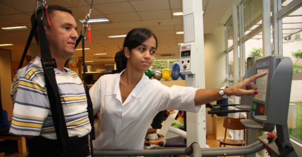

During FAPESP Week Michigan-Ohio, a researcher from Brazil presented methodology used to stimulate neuroplasticity and improve motor function (photo: robotic exoskeleton/ release)
During FAPESP Week Michigan-Ohio, a researcher from Brazil presented methodology used to stimulate neuroplasticity and improve motor function.
During FAPESP Week Michigan-Ohio, a researcher from Brazil presented methodology used to stimulate neuroplasticity and improve motor function.

During FAPESP Week Michigan-Ohio, a researcher from Brazil presented methodology used to stimulate neuroplasticity and improve motor function (photo: robotic exoskeleton/ release)
By Karina Toledo, in Michigan | Agência FAPESP – A number of innovative techniques, which includes robotic exoskeletons, transcranial magnetic stimulation (TMS) and high-density electroencephalography (HD-EEG) have been used successfully by researchers at the Physical and Rehabilitation Medicine Institute (IMREA) of the University of São Paulo School of Medicine (FMUSP) in treating patients who suffer from cerebrovascular accident (CVA) and spinal cord injury.
Findings of the research study were presented in the U.S. by Professor Linamara Rizzo Battistella March 28, 2016 during FAPESP Week Michigan-Ohio. The event, which runs through April 1, was organized by the São Paulo Research Foundation (FAPESP) together with the University of Michigan (UM) and Ohio State University (OSU) for the purpose of promoting increased cooperation between scientists from São Paulo and the United States.
Some of the data were also recently published in an article in the journal Restorative Neurology and Neuroscience.
“Our group has two main objectives: identify each patient’s potential for motor recovery and, once this objective has been achieved, look for ways in which this individual can engage in the everyday activities of life with appropriate adaptations such as by using a walker or a wheelchair,” Battistella explained in an interview given to Agência FAPESP.
One of the key points in caring for patients suffering from CVA is identifying predictors of motor recovery – signals captured by recording the brain’s electrical activity that indicate each patient’s capacity to regain movement.
This is done by using a combination of two techniques: monitoring motor evoked potential (MEP) – a test that applies magnetic stimulation to the brain and evaluates motor response – and measuring the electrical activity of the brain using HD-EEG.
"MEP identifies what we call the motor threshold, which is an objective measurement of the potential for motor recovery,” the researcher explained.
The neurophysiological approach also includes using transcranial magnetic stimulation for diagnostic purposes as a way to indicate which areas of the brain need to be stimulated or inhibited in order to induce neuroplasticity and improve motor control.
According to Battistella, both brain stimulation as well as inhibition are done using a device that applies repetitive transcranial magnetic stimulation pulses (rTMS), which is different from what is used to map brain activity. The method’s goal is to promote balance in the activity of the two hemispheres of the brain.
In parallel with the neurophysiological evaluation, clinical tests are run in which patients are asked to conduct a series of predetermined movements that are then assessed on a scale such as Fugl-Meyer. At the end, each patient receives a score according to what she has been able to accomplish.
The data from clinical and neurophysiological evaluations are then statistically analyzed. “This way we are able to objectively determine the state of recovery of the injured hemisphere and plan appropriate treatment,” the researcher said.
“We compared a group of patients undergoing only a conventional rehabilitation program with a group of patients who, in addition to the exercises, received magnetic stimulation to promote cortical stability. This second group presented considerable improvement over the first. We can therefore say that the technique is able to indirectly influence the processes of neuroplasticity and thus, improve motor control.
In terms of the robotic exoskeleton, in addition to helping in walking or moving one’s arms, it also provides researchers with objective measurements of each patient’s functional performance. Using sensors applied to the upper and lower limbs, the device calculates how much force an individual puts forth during the movements. The data are then sent to a computer and displayed as graphs.
“The patient is able to see improvement in every session and realize that she depends less and less on help from the device to walk or move her arms. This provides positive reinforcement, improves performance and increases treatment adherence,” Battistella said.
According to the researcher, all these related measures allow recognition of the biomarkers for brain plasticity in patients with brain injury. In other words, they help scientists understand how the brain is working after the injury and how brain reorganization is taking place.
"Neuroplasticity is the brain’s ability to reorganize itself after an injury, strengthening the neural networks that were not affected and ensuring an adequate level of functionality. The successful outcome of rehabilitation depends upon this process of reorganization. Our intent, based on test results, is to make treatment more effective and shorten the time it takes for rehabilitation,” Battistella explained.
The group is working together with Harvard Medical School’s Laboratory of Neuromodulation lead by Professor Felipe Fregni. One of the principal investigators at IMREA-FMUSP is Professor Marcel Simis.
Spinal cord injury
The cases of paraplegia and quadriplegia at the Hospital das Clínicas at the USP School of Medicine mainly involve young males between the ages of 17 and 30.
“From the beginning, this group has presented a more clearly-defined prognosis. The capacity for motor recovery is directly related to the severity and location of the injury. Our role in this case, besides identifying and developing the motor potential for each individual, is to prevent the secondary complications of these conditions,” Battistella said.
Individuals with disabilities resulting from spinal cord injury, the researcher said, frequently suffer from urinary infections, kidney failure, osteoporosis and bedsores, in addition to sarcopenia (loss of muscle mass and strength) and joint deformities that can aggravate the functional condition of these patients over the years.
“Robotic walking, for instance, can help prevent osteoporosis because it stimulates bone metabolism. Magnetic stimulation as well as that which uses electrical current prevents atrophy of the brain regions that no longer receive motor stimuli as a result of the injury. Stimulation in other regions of the brain may in turn offer increased motor function,” the researcher said.
Neuromodulation: other applications
Another aspect of the collaboration between IMREA and Harvard combines transcranial direct current stimulation (TDCS) with aerobic exercise as a way to treat chronic pain in individuals who suffer from fibromyalgia. The method can also be used on patients who suffer chronic pain from incomplete spinal cord injury.
"We use a device during exercise that produces electrical stimuli that are well-tolerated by the brain and have the ability to control pain. The current inhibits the area that modulates the phenomenon of pain. It is as if we were administering an analgesic,” she said.
Republish
The Agency FAPESP licenses news via Creative Commons (CC-BY-NC-ND) so that they can be republished free of charge and in a simple way by other digital or printed vehicles. Agência FAPESP must be credited as the source of the content being republished and the name of the reporter (if any) must be attributed. Using the HMTL button below allows compliance with these rules, detailed in Digital Republishing Policy FAPESP.


