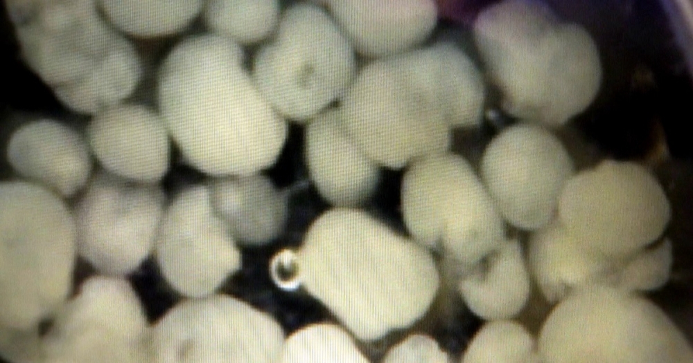

Model developed for research into neuropsychiatric disorders is being used to investigate effects of Zika virus on the brain and to pursue therapies (image: mini-brains/IDOR Lab Team)
Model developed for research into neuropsychiatric disorders is being used to investigate effects of Zika virus on the brain and to pursue therapies.
Model developed for research into neuropsychiatric disorders is being used to investigate effects of Zika virus on the brain and to pursue therapies.

Model developed for research into neuropsychiatric disorders is being used to investigate effects of Zika virus on the brain and to pursue therapies (image: mini-brains/IDOR Lab Team)
By Karina Toledo | Agência FAPESP – Brazilian scientists have used cerebral organoids – three-dimensional structures called mini-brains that are created in a laboratory from induced pluripotent stem cells (iPSCs) – to understand the link between infection with Zika virus and the development of microcephaly.
Part of their research, including experiments led by Stevens Rehen and Patrícia Garcez at the Federal University of Rio de Janeiro’s Institute of Biomedical Sciences (ICB-UFRJ) and the D’Or Institute for Research and Education (IDOR), also in Rio de Janeiro, has just been published in Science. Juliana Minardi Nascimento also participated, with support from FAPESP via a postdoctoral scholarship.
“My postdoctoral project is about using brain organoids generated from cells supplied by patients with schizophrenia to do proteomics, which means evaluating all the proteins expressed in these cells and comparing them with the proteins expressed in cells of donors without the disease to understand what’s different. The main advantage of this model is that it leaves intact the genetic information provided by the patient,” Nascimento said.
Affiliated with the Neuroproteomics Laboratory at the University of Campinas’s Biology Institute (IB-UNICAMP) and working under the supervision of Professor Daniel Martins de Souza, Nascimento decided to collaborate with the Rio de Janeiro group, one of the few in the world to have mastered the technique of creating mini-brains to study neuropsychiatric diseases.
The technique’s first step is to reprogram ordinary mature cells (which may be skin cells or bladder epithelial cells taken from urine) so that they become as pluripotent as embryonic stem cells. The 2012 Nobel Prize in Medicine was awarded to Shinya Yamanaka of Kyoto University in Japan for inventing this procedure (in 2006).
These iPSCs are then induced by chemical stimuli to differentiate into neural stem cells, a type of progenitor cell that can self-renew to generate several types of brain cell, such as neurons, astrocytes and microglia.
Mini-brains are grown by making this process occur three-dimensionally under specific conditions in the lab. “Instead of a culture plate, the cells are put into a rotating culture system along with certain other elements,” Nascimento said. “Cell communication changes as they become three-dimensional. In a two-dimensional culture, each cell interacts with its immediate neighbor, whereas in the 3D model, communication takes place with all surrounding cells. They function very similarly to cells in the developing brain.”
Thus, in the right conditions, neural stem cells organize themselves into layers, initially forming neurospheres and then cerebral organoids. The former mimic the brain of a rudimentary embryo, while the latter are comparable to the brain of a three-month-old fetus.
Nascimento plans to develop the models for the study of schizophrenia in Rio de Janeiro and then perform proteomics analysis at UNICAMP. Since the emergence of the epidemic caused by Zika virus, she has been pursuing this research in parallel with studies designed to understand the effects of infection on the brain.
“We knew cerebral organoids would be useful in our efforts to understand the link between Zika and microcephaly,” she said. “This is an important public health issue, so part of Rehen’s lab team decided to concentrate on that target. The work has developed very quickly.”
Three different experiments were performed to test Zika’s effects on the brain. The first step was to infect two-dimensionally cultivated neural stem cells. The group found that the virus caused cell death in about three days.
In a second experiment, the stem cells were infected and induced to differentiate into the 3D model. After three days, Rehen and his team found that the cells’ ability to generate neurospheres had been crippled by Zika.
“A few neurospheres were formed, but they degraded in up to six days, whereas those originating from uninfected cells developed normally,” Nascimento said.
Electron microscope images showed that the virus had multiplied rapidly inside the cells, giving rise to a process of programmed cell death called apoptosis.
In a third experiment, 30-day-old mini-brains were infected with Zika and, when compared with uninfected organoids after 11 days, were found to have grown 40% less than the controls.
“Although the mini-brains weren’t destroyed like the neurospheres, their growth was clearly impaired. We’re now investigating whether cell differentiation was also affected in the model,” Nascimento said.
Next steps
In partnership with researchers at UNICAMP, the Rio de Janeiro group plans to perform proteomic analysis of the mini-brains affected by Zika to identify which biochemical pathways are changed by the infection. The answer could suggest possible therapies.
“Our goal is to see whether there are any existing drugs approved for human use that can mitigate the impact of the virus on brain cells,” Nascimento said. “We’re already testing some compounds, from food supplements to antivirals. One of these looked promising in preliminary tests.”
Republish
The Agency FAPESP licenses news via Creative Commons (CC-BY-NC-ND) so that they can be republished free of charge and in a simple way by other digital or printed vehicles. Agência FAPESP must be credited as the source of the content being republished and the name of the reporter (if any) must be attributed. Using the HMTL button below allows compliance with these rules, detailed in Digital Republishing Policy FAPESP.


