


The methodology was developed in the United States with the help of a Brazilian researcher. The therapeutic approach was tested on carriers of hemophilia (image: Wikimedia)
The methodology was developed in the United States with the help of a Brazilian researcher. The therapeutic approach was tested on carriers of hemophilia.
The methodology was developed in the United States with the help of a Brazilian researcher. The therapeutic approach was tested on carriers of hemophilia.
The methodology was developed in the United States with the help of a Brazilian researcher. The therapeutic approach was tested on carriers of hemophilia (image: Wikimedia)
By Karina Toledo, in Campinas
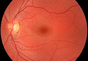
Agência FAPESP – More than 20 carriers of a type of blindness caused by a genetic defect known as Leber’s congenital amaurosis (LCA) have recovered their sight thanks to a treatment developed by researchers at the University of Pennsylvania and the Children’s Hospital of Philadelphia in the United States.
One of the leaders of the group is Brazilian Valder Arruda, who presented the results of the tests during the FAPESP-sponsored event “Advanced Topics in Genomics and Cell Biology,” which was held August 4-6, 2014, at the University of Campinas (Unicamp).
“We have demonstrated the safety and effectiveness of the treatment and are now at the stage of finding the dose capable of having a therapeutic effect on patients. I think this may be the first gene therapy approved for commercial use by the FDA [the U.S. Food and Drug Administration]. It has taken many years of study and testing to get to this stage,” said Arruda, former Unicamp professor and current faculty member at the University of Pennsylvania.
In an interview with Agência FAPESP, the researcher explained that we know of at least 15 genes that, when mutated, result in congenital blindness such as LCA. All mutations cause progressive degeneration of the retina that begins at birth.
“From very early on, the children’s vision is compromised, and ocular movements are unusual. They generally end up blind by adolescence,” Arruda explained.
The therapy developed by the group at the University of Pennsylvania, that also includes leadership from ophthalmologist Jean Bennett, is designed to treat carriers of a mutation in the gene that encodes the RPE65 protein, which is involved in the metabolism of vitamin A, an essential nutrient for eye tissue health.
“We decided to begin the studies using the RPE65 gene because some dogs have the same mutation that is often found in humans, which makes it easier to conduct preclinical testing. The technology can be adapted for other forms of congenital amaurosis, and there are groups in several countries that are conducting research with this goal in mind. Of course, every case is different,” Arruda said.
The method consists of inserting a healthy copy of the RPE65 gene within a modified virus. The viral vector selected by the researchers was the adeno-associated virus (AAV), a type of parvovirus that does not cause disease in humans.
“We removed the genetic material from the AAV and inserted a copy of the RPE65 and a promoter – the molecule that will send a message to activate gene expression – in its place. The idea is to take advantage of the natural capacity of the virus to infect human cells and deposit the genetic material in them. That way, the cells of the retina can begin to express the RPE65 protein that was previously deficient,” the researcher explained.
In 2001, Bennett’s group published promising results of tests performed on dogs in the journal Nature Genetics, and then, in partnership with Arruda, initial trials on humans began.
“In phase 1 clinical trials, the objective was to ensure the safety of the treatment. We injected a low dose of the viral vector into three adult carriers of the mutation. Normally, phase 1 trials are only performed on healthy patients, but it’s different in the case of gene therapy,” Arruda explained.
Initial exams conducted after treatment revealed that pupils that had previously not reacted to light began to respond in almost the same way as those of an individual with normal vision.
“Three weeks after applying the viral vector, one of the patients said she was able to make out the shape of the furniture in her apartment. At first I thought this was only a placebo effect, but a few weeks later, another patient reported something similar,” Arruda said.
Later tests carried out by an independent team confirmed that the three volunteers had recovered partial vision, contradicting the prevailing notion in the field of neuroscience that the brains of individuals who had spent many years without seeing would have totally lost the capacity to interpret the signs captured by the retina.
“This first Italian patient was 36 years old. Using functional magnetic resonance exams, we showed that the areas of the visual cortex that had been inactive before the treatment began to show activity 30 days after injection with the viral vector. And there was even more activity 90 days later. These results could potentially question the prevailing dogma of neuroscience,” Arruda said.
The initial trials conducted on 12 patients, including an 8-year-old boy, were published in 2009 in the journal The Lancet and showed that the effectiveness of treatment was directly related to its early start.
“Normally, only adults over age 18 are allowed to take part in phase 1 trials. But because this is a degenerative disease, there may not have been any retinal tissue left to restore if we had waited much longer to begin the treatment. Because of this, we received authorization to include children. Now, we’re beginning phase 3 trials, and we’ve lowered the minimum age to 4,” Arruda said.
Until now, 24 patients from countries such as Italy, the Netherlands, Belgium, the United States and Australia have received intraocular injection of the viral vector – first in one eye and then in the other – and have presented with better vision, with no signs of toxicity or immunological reaction.
“We have a 9-year-old girl who has begun to play hide and seek, a 16-year-old boy who was able to get his driver’s license, and many adults who have become independent. All are feeling more confident,” Arruda said with pleasure.
Hemophilia
Before joining Bennett’s team in treating retinal diseases, Arruda, a hematologist by training, had been testing a similar methodology to treat one of the forms of hemophilia, a genetic disease characterized by problems with blood clotting.
“There are two types of hemophilia: A and B. The first occurs because of a deficiency of a protein known as clotting factor VIII, and the second occurs due to a deficiency of clotting factor IX. Suitable levels of both proteins are needed in the plasma so that blood clotting works correctly when necessary. However, when either of the factors is deficient, the manifestation is the same,” Arruda explained.
The disease is considered mild and may not be perceived for many years if the deficient clotting factor – VIII or IX – still makes up over 5% of the clotting factors that are normally found in the blood of individuals who are not hemophiliacs. When the level is between 1% and 5%, the disease is considered to be moderate; below 1% is considered severe. In these cases, bleeding may occur spontaneously, even without significant trauma.
“The more severe it [the disease] is, the more frequent the complications, such as bleeding in the joints, muscles or intracranially. Patients with severe disease generally bleed once a month. Moderate patients bleed two or three times every six months. Mild cases may go an entire year without bleeding,” Arruda said.
The treatment that is available today, explained the researcher, involves replacing the deficient clotting factor, but the protein only lasts for a short time before needing to be replaced. The therapy may be performed routinely as a preventative measure in the most severe cases; however, the main constraint is the cost, which could exceed US$200,000 per year for a child between the ages of 10 and 15.
“In Brazil, the Unified Health System (SUS), in most cases, offers the clotting factor only when the patient is already bleeding or in cases where the patient requires a surgical procedure. But every time there is bleeding, the region becomes more compromised and will bleed more easily the next time. The ideal is prophylactic treatment,” Arruda said.
To obtain a curative method, over 15 years ago, the researchers at the University of Pennsylvania began to develop a modified AAV with a copy of the gene responsible for producing factor IX. The tests and trials went through various phases.
“Although hemophilia B is not the most prevalent form, we began with that gene because it is much smaller than the factor VIII gene and would be easier to insert into the viral vector. Now, we’re developing a version of the therapy for hemophilia A. The goal is to continually maintain the levels of blood clotting factor above 1% of the normal level to reduce the need for replacement treatment. If we are able to keep these levels above 6%, the patient would only need to replace the protein if undergoing surgery,” Arruda noted.
The initial tests conducted on a lineage of dogs that carried severe hemophilia type B were published in 2002 in the journal Blood. The animals were monitored for two years and, after a single injection, maintained stable plasma factor IX levels between 6% and 9%. They are still considered to be carriers of mild hemophilia.
At the same time, the first clinical trials began with seven adult volunteers, and for the first time, a viral vector was introduced to the human liver through the hepatic artery using a catheter.
Although the approach has proven to be safe, to the surprise of the researchers the plasma factor IX levels that had increased shortly after application of the treatment began to fall a few weeks later.
New studies have revealed that the liver cells that had begun to produce factor IX thanks to the gene therapy were being destroyed by the immune system, which recognized the viral vector as a foreign body. This effect had not been observed in dogs and was it seen later in monkeys.
“It’s possible that these patients had already been infected by AAV and had created antibodies against the proteins of the viral envelope,” Arruda said.
The results of this first clinical test were published in 2006 in the journal Nature Medicine.
In partnership with other colleagues from the Children’s Hospital of Philadelphia, a group from St. Jude Children’s Research Hospital, also in the United States, carried out new phase 1 clinical trials. This time, researchers closely monitored the changes in the hepatic enzyme and factor IX levels in the blood.
At the slightest sign that the hepatocrits were being destroyed by the immune system, an immunosuppressive drug was administered. The strategy worked, and the results were published in 2011 in The New England Journal of Medicine.
“The immunosuppressive drug was used for only eight weeks, because over time, the proteins of the viral envelope are degraded and the immune system stops attacking the hepatocrits. One of the patients maintained 6% to 8% of the normal factor IX level in the plasma, while another, who had taken more time to achieve immunosuppression, fell to 3%. We needed to act quickly,” Arruda said.
Even in cases in which the levels remained between 1% and 2% of the normal level, the researcher added, a 77% reduction in the need to replace the protein has been possible.
The Agency FAPESP licenses news via Creative Commons (CC-BY-NC-ND) so that they can be republished free of charge and in a simple way by other digital or printed vehicles. Agência FAPESP must be credited as the source of the content being republished and the name of the reporter (if any) must be attributed. Using the HMTL button below allows compliance with these rules, detailed in Digital Republishing Policy FAPESP.
