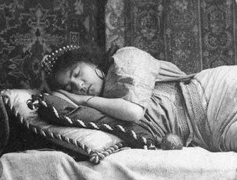

Study shows that the integrated neural respiratory control system makes expiration cease to be passive when ambient carbon dioxide increases (photo: Charles Ellis Johnson / Wikimedia)
Study shows that the integrated neural respiratory control system makes expiration cease to be passive when ambient carbon dioxide increases.
Study shows that the integrated neural respiratory control system makes expiration cease to be passive when ambient carbon dioxide increases.

Study shows that the integrated neural respiratory control system makes expiration cease to be passive when ambient carbon dioxide increases (photo: Charles Ellis Johnson / Wikimedia)
By Maria Fernanda Ziegler | Agência FAPESP – Breathing consists of pulmonary ventilation via inspiration, the inhalation of air to provide oxygen, and expiration, the exhalation of spent air to remove carbon dioxide. Inspiration is a spontaneous but active process that involves the contraction of a group of muscles including the diaphragm. Expiration is normally passive, involving only lung and muscle recoil.
Hypercapnia is a condition that occurs when there is too much carbon dioxide in the bloodstream, typically owing to inadequate respiration. It tends to make expiration become active by recruiting the abdominal muscles, among others, to increase the exhaled volume. Hypercapnia-induced active expiration increases automatically during, according to a study by researchers at São Paulo State University’s School of Agricultural & Veterinary Sciences (FCAV-UNESP) in Jaboticabal, Brazil.
“We succeeded in showing that when ventilatory demand increases in hypercapnia, the abdominal muscles go into action, and expiration ceases to be passive. This recruitment of the abdominal muscles in hypercapnia occurs whether the individual is awake or asleep, but it’s more intense during sleep,” Glauber dos Santos Ferreira da Silva, one of the authors of the study, told Agência FAPESP.
The study was supported by FAPESP through its Young Investigator Grant Program and two scientific initiation scholarships and was published in The Journal of Physiology.
Male adult rats were exposed for 60 minutes to a hypercarbic gas mixture analogous to hypercapnia (7% CO2) while their brains were monitored by EEG and their diaphragm and abdominal muscles by EMG. The data showed that under hypercapnia conditions, active expiration – respiratory recruitment of the abdominal muscles – was predominant during non-rapid-eye-movement (REM) sleep.
Da Silva explained that the analysis during sleep was restricted to non-REM periods because the characteristics of sleep in rats are different from those of sleep in humans.
“Rats are most active at night and sleep during the day. All our experiments took place in daytime. Also, rats don’t sleep continuously, as most humans do. They go through the same stages as we do but in a more fragmented manner, with very short REM periods that can’t properly be captured during hypercapnia,” da Silva said.
The study found that abdominal muscle activity was intense and became more variable during sleep than during waking hypercapnia periods. Similarly, active expiration was much more prevalent during sleep, albeit intermittent.
“This is probably the case in humans as well,” da Silva said. “The process of abdominal muscle recruitment has been studied since the 1980s, but interest in this research field has revived in the last five years, since the discovery of a region of the central nervous system called the parafacial respiratory group that’s responsible for originating this expiratory activity.”
Hypercapnia and obstruction
Hypercapnia is frequent in patients with airway obstruction conditions, of which obstructive sleep apnea is the most familiar. In these conditions, loss of muscle tone in the upper airway muscles, which control airway dilation and wall stiffening, leads to airway collapse.
These individuals display hypercapnia during sleep. Respiration stops, and CO2 builds up in the bloodstream instead of being expelled. “There are reports of abdominal muscle recruitment in humans with sleep hypercapnia, which is precisely the condition to which we exposed the rats during our study,” da Silva said.
In the study, exposure to hypercapnia caused the partial pressure of CO2 in the rats’ bloodstream to increase by 15 mmHg, which is similar to what occurs in humans with obstructive sleep apnea. In these situations, “appropriate respiratory motor outputs, as well as ventilatory adjustments to hypercapnia, are highly desirable,” the authors explain in the article.
For da Silva, the findings are relatively simple but represent important advances. “One of these relates to the perspective of neural control of breathing,” he said. “We didn’t set out to investigate neural mechanisms, but we used a model and methods with high integrative value in the physiology of respiratory control. A question that remains is this: given that neurons are producing abdominal activity during sleep, some mechanism must be responsible for activating these neurons in the central nervous system. Understanding such mechanisms is a challenge that is still posed in the literature. The continuation of this study will set out to investigate the possible neural mechanisms that are involved in this kind of response.”
A commentary on the study was published in the “Perspectives” section of the same issue of The Journal of Physiology.
“The finding of increased active expiration during restful non-REM sleep is intriguing in the context of a generalized decrease in upper airway muscle activity, but not the diaphragm, during sleep with relevance to obstructive airway events characteristic of obstructive sleep apnea syndrome,” writes Ken D. O’Halloran, a professor in University College Cork Medical School’s Department of Physiology in Ireland, who did not participate in the study.
According to O’Halloran, hypoventilation “may provide additional reflex-mediated impetus to active expiration during sleep, perhaps defending against overt repression of breathing.”
And he goes on to say, “Studies of the expiratory phase of the respiratory cycle have returned to center stage and have never been so active. The technically challenging study by Leirão et al. offers an integrative perspective on brainstem oscillatory models of respiratory control, unmasking the pattern of abdominal muscle recruitment during respiratory challenge to reveal the mechanical consequences of such behavior for pulmonary ventilation.”
The article “Hypercapnia-induced active expiration increases in sleep and enhances ventilation in unanesthetized rats” (doi: 10.1113/JP274726) by Isabela P. Leirão, Carlos A. Silva Jr, Luciane H. Gargaglioni and Glauber S. F. da Silva can be retrieved from onlinelibrary.wiley.com/doi/10.1113/JP274726/full.
Republish
The Agency FAPESP licenses news via Creative Commons (CC-BY-NC-ND) so that they can be republished free of charge and in a simple way by other digital or printed vehicles. Agência FAPESP must be credited as the source of the content being republished and the name of the reporter (if any) must be attributed. Using the HMTL button below allows compliance with these rules, detailed in Digital Republishing Policy FAPESP.


