


Current tests at a Brazilian institution have developed optical coherence tomography to enable virtual biopsy of lesions (A.C. Camargo Cancer Center)
Current tests at a Brazilian institution have developed optical coherence tomography to enable virtual biopsy of lesions.
Current tests at a Brazilian institution have developed optical coherence tomography to enable virtual biopsy of lesions.
Current tests at a Brazilian institution have developed optical coherence tomography to enable virtual biopsy of lesions (A.C. Camargo Cancer Center)
By Karina Toledo
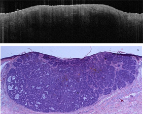
Agência FAPESP – A new technology known as optical coherence tomography (OCT) promises to make diagnosing skin cancer much more precise and much less invasive. Currently, in tests conducted at the A.C. Camargo Cancer Center, this method has been applied for the virtual biopsy of lesions, to confirm malignancy in cases of doubt and to help define the margins and the characteristics of the tumor, which represent important factors in creating a plan for treatment.
The OCT equipment, which was acquired with the support of FAPESP, is similar to that used for ultrasound. Instead of sound waves, however, this approach emits a low-frequency laser – roughly 1,300 nanometers – capable of producing a high-resolution three-dimensional image.
“This technique allows us to conduct similar analyses as those performed for histopathological exams of biopsy tissues and is currently considered the gold standard in skin cancer diagnosis,” explained dermatologist Gisele Gargantini Rezze, who coordinates the experiments in partnership with pathologist Maria Dirlei Begnami.
According to Rezze, there are other methods to obtain auxiliary images in skin cancer, such as dermatoscopy – a type of digital magnifying glass that amplifies the lesion by 20 times and is widely used in the daily routine of dermatology offices.
There is also focal microscopy, consisting of experimental equipment that was acquired by the A.C. Camargo Cancer Center with FAPESP financing, and this approach uses a 850-nanometer laser to magnify the lesion up to 900 times.
“The resolution of confocal microscopy is even greater than that of optical coherence, although this equipment has major limitations because it does not provide information on the depth of the lesion,” explained Rezze.
OCT also offers other advantages, explained the researcher. The exam last just two minutes and produces images in real time and in transversal cuts with up to 2.5 millimeters in depth, which is the equivalent of the deepest portion of the dermis. In contrast, assessment with confocal microscopy is more laborious, as this approach requires roughly 40 minutes and generates only images in horizontal cuts, reaching a maximum depth of 1.5 millimeters.
“Sometimes the epidermis – the most superficial part of the skin – is normal and the tumor is located in the dermis. In the cases of recidivism, for example, it is common for the tumor to reappear below the first surgical scar. In these situations, OCT can better evaluate the lesion and help the doctor prepare for surgery because it offers information on the exact location of the tumor and its margins,” said Rezze.
The exam, however, is not for all patients, warned the researcher. Because the equipment is expensive – costing R$130,000 – it is recommended mainly for the evaluation of questionable lesions when the doctor cannot confirm the malignancy through other diagnostic imaging techniques.
“I believe that the major advantage of this technology will be a reduction in unnecessary surgeries. This represents savings of time and money for both patients and health centers,” evaluates Rezze.
New parameters
Before being incorporated into the medical routine, however, new equipment must be studied and familiarized. Furthermore, its diagnostic accuracy must be validated. One of the objectives of the study coordinated by Rezze is establishing parameters that will help doctors to interpret images generated by OCT.
“We need to learn to read the results and distinguish between melanoma and normal moles, for example. For this reason, we will describe different types of lesions in different anatomical regions and compare them to histopathological exams and dermatoscopy,” explained Rezze.
According to the researcher, 15 centers around the world are currently testing cancer diagnosis using OCT. This technology has also been used for the evaluation of ophthalmological disturbances.
The Agency FAPESP licenses news via Creative Commons (CC-BY-NC-ND) so that they can be republished free of charge and in a simple way by other digital or printed vehicles. Agência FAPESP must be credited as the source of the content being republished and the name of the reporter (if any) must be attributed. Using the HMTL button below allows compliance with these rules, detailed in Digital Republishing Policy FAPESP.
