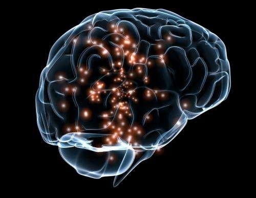

In experiments with animals, a Cleveland Clinic research group shows that DBS assists formation of new synapses and neurons, bolstering motor rehabilitation (image: Wikimedia Commons)
In experiments with animals, a Cleveland Clinic research group shows that DBS assists formation of new synapses and neurons, bolstering motor rehabilitation.
In experiments with animals, a Cleveland Clinic research group shows that DBS assists formation of new synapses and neurons, bolstering motor rehabilitation.

In experiments with animals, a Cleveland Clinic research group shows that DBS assists formation of new synapses and neurons, bolstering motor rehabilitation (image: Wikimedia Commons)
By Karina Toledo | Agência FAPESP – Deep brain stimulation (DBS), already used in humans to treat symptoms of Parkinson’s disease, is being tested to aid post-stroke recovery from paralysis.
Studies in animal models were led by Andre Machado, Brazilian-born chairman of the Neurological Institute at Cleveland Clinic, Ohio, in the United States, and director of the Clinic’s Center for Neurological Restoration. The team has applied to the US health authorities for permission to begin first-in-human studies, initial clinical trials in which experience with animal testing is extended to human subjects for the first time.
DBS involves surgically implanting very fine wires with electrodes at their tips into deep areas of the brain. A pulse generator similar to a pacemaker is placed under the skin near the collarbone. The device delivers electrical impulses to the brain, modulating the activity of the target nerve structure and stimulating the formation of new synapses. The technique, which may even help create new neurons, is being studied by other groups as an aid in the treatment of depression and chronic pain.
“In the case of Parkinson’s, the technique mitigates some of the main symptoms, including tremor, stiffness, and slow movement. In trials with laboratory animals, we found that DBS significantly improves outcomes in post-stroke physical rehabilitation,” Machado told Agência FAPESP.
Using an animal model, the group set out to stimulate the dentate nucleus, a region of the cerebellum that has extensive direct connections with the cortex.
The researchers used two techniques to induce ischemia in rats. The first consisted of an intracortical injection of endothelin, a drug that reduces the flow of blood through the middle cerebral artery. In the second technique, the artery was coagulated and dissected by microsurgery. In both cases, infarction was induced in the region irrigated by the artery, similarly to what occurs when the artery is occluded by atherosclerosis. The death of brain tissue in this region normally results in partial paralysis on the opposite side.
A pulse generator for DBS was then implanted in each of the animals, and they were submitted to a period of rehabilitative training similar to physical therapy. Half underwent DBS. The device was not activated in the other half, which served as a control group.
The researchers compared motor recovery between the control group, which was only subjected to physical training, and the group that received DBS as well as training.
“We measured the improvement by means of tasks that are well defined in the scientific literature,” Machado said. “These are designed to encourage the animals to use the stroke-affected paw to reach for bits of food and put them in their mouths. The number of pieces retrieved can be compared in order to create a motor function recovery index. The group subjected to DBS performed significantly better than the control group.”
Action mechanism
When they investigated the mechanisms through which the therapy produced an improvement, the team discovered that the treated animals had twice as many synapses in the stroke-affected area compared with the control group.
They also observed an increase in the expression of proteins related to long-term potentiation (LTP), a phenomenon associated with brain plasticity processes.
“These findings suggest therapy assists reorganization of the brain so that other regions can take over some of the functions previously performed by the affected areas,” Machado said.
Studies conducted more recently by the group and not yet published indicate that therapy also induces neurogenesis, the process of new neuron formation, in stroke-affected areas.
“We used a method called immunohistochemistry to analyze samples of brain tissue from the rats submitted to treatment and found a statistically significant increase in the number of new cells compared with the control group,” Machado said.
Some of the results obtained thus far have been published in articles in Brain Stimulation, The Journal of Neuroscience, and Frontiers in Systems Neuroscience.
In the US, some 800,000 people survive a stroke every year. Only 10% recover almost completely, while 15% die soon afterward. Approximately 25% are left with mild disability, but 50% are severely disabled and require special care.
According to the Brazilian Cardiology Society (SBC), strokes cause 100,000 deaths every year in Brazil, affecting men and women in very similar proportions (50.5% and 49.5%, respectively). The SBC estimates that 300,000 people survive a stroke annually, some of whom suffer disability as a result.
Republish
The Agency FAPESP licenses news via Creative Commons (CC-BY-NC-ND) so that they can be republished free of charge and in a simple way by other digital or printed vehicles. Agência FAPESP must be credited as the source of the content being republished and the name of the reporter (if any) must be attributed. Using the HMTL button below allows compliance with these rules, detailed in Digital Republishing Policy FAPESP.


