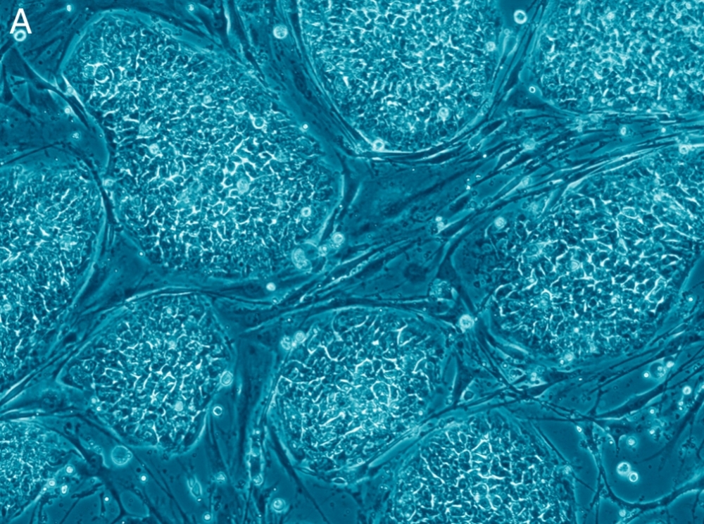

In studies with tumor stem cells, researchers at Uruguay’s University of the Republic have identified regulators with the potential to interfere in the development of cancer cells (image: Wikimedia Commons)
In studies with tumor stem cells, researchers at Uruguay's University of the Republic have identified regulators with the potential to interfere in the development of cancer cells.
In studies with tumor stem cells, researchers at Uruguay's University of the Republic have identified regulators with the potential to interfere in the development of cancer cells.

In studies with tumor stem cells, researchers at Uruguay’s University of the Republic have identified regulators with the potential to interfere in the development of cancer cells (image: Wikimedia Commons)
By Heitor Shimizu, in Montevideo | Agência FAPESP – Studies indicate the presence of tumor stem cells in solid tumors and blood cancers such as leukemia, myeloma and lymphoma. Their characteristics are similar to those of normal stem cells, especially the capacity to originate any of the cell types found in the various forms of cancer.
Tumor stem cells, also known as cancer stem cells, can divide, self-renew and give rise to all cell types found in tumors. For this reason they are an important target for researchers in several countries. However, the generality of the cancer stem cell thesis has also been challenged by scientists who do not believe such cells exist or that normal stem cells can give rise to cancer stem cells. Others more cautiously prefer to say their existence is still hypothetical.
The cells found in malignant tumors are not always the same. Tumors may contain a variety of cell types, and the idea of tumor stem cells is that some cancer cells always act like stem cells that reproduce and sustain the cancer, similarly to the way in which normal stem cells become organs and tissue.
A group of researchers at Uruguay’s University of the Republic (UDELAR) has no doubts about the existence of cancer stem cells and wants more research to be done on the subject.
“Tumor or cancer stem cells are extremely interesting from a clinical standpoint because they’re involved in metastasis and drug resistance, impairing treatments such as chemotherapy,” said Maria Ana Duhagon, a researcher affiliated with UDELAR Medical School’s Molecular Interaction Laboratory.
Several kinds of cancer therapy are designed to reduce tumor size but the patient may suffer a relapse if the treatment does not destroy the tumor stem cells.
At FAPESP Week Montevideo, held on November 17-18 in Uruguay, Duhagon spoke about her group’s research with microRNAs involved in stem cell differentiation in prostate cancer, the second most frequent cause of death for men in Uruguay and Brazil.
MicroRNAs are non-coding RNAs (ribonucleic acids) that play a key role in the regulation of gene expression in plants and animals, generally remaining in the cell nucleus. Non-coding RNA is transcribed from DNA and not translated into polypeptides.
“We’ve developed a method of isolating, maintaining and differentiating tumor stem cells that can be used to analyze sensitivity to conventional and natural drugs, and to identify pathways that modulate differentiation,” Duhagon said. “The method has also enabled us to determine metastasis capacity and to characterize microRNAs modulated during differentiation.”
MicroRNAs are known to be largely downregulated in cancer, enabling them to be used to identify or monitor the development of the disease.
“MicroRNAs participate in maintaining the state of tumor cell differentiation. This is our group’s fundamental line of research,” Duhagon said.
Duhagon and her group have identified microRNAs with therapeutic potential for the treatment of prostate cancer. By detecting possible sites for interaction between one of these miRNAs (hsa-miR-886-3p) and messenger RNA in the three-prime untranslated region (3’UTR), they were able to select several candidate tumor suppressor genes.
“These genes’ messenger RNA diminished with the temporary transfer of hsa-miR-886-3p, inhibiting proliferation,” Duhagon said.
“Hsa-miR-886-3p comes from an unconventional RNA precursor designated vtRNA2-1 and located at a site of emerging relevance but poorly understood. Expression of hsa-miR-886-3p and vtRNA2-1, which we found in samples obtained in Uruguay and from the Cancer Genome Atlas, is strongly regulated by methylation of their promoter, making it a possible tumor suppressor gene in prostate cancer.” Methylation is a chemical reaction that adds methyl groups to RNA and DNA, regulating gene expression.
Their analysis identified the tumor suppressor’s role in the G2/M phase, affecting proliferation in both in-vitro assays and animal models. G2/M is the mitotic entry checkpoint, an important phase of the cell cycle that checks for DNA damage.
“We used gene expression microarrays to identify 278 transcriptions regulated differently by vtRNA2-1, representing cellular processes such as cell migration, cell adhesion and cell cycle,” Duhagon said. A microarray is a set of DNA sequences representing hundreds or thousands of genes arranged in a grid pattern for use in genetic testing of samples and measurement of gene expression levels.
Read more about FAPESP Week Montevideo: www.fapesp.br/week2016/montevideo.
Republish
The Agency FAPESP licenses news via Creative Commons (CC-BY-NC-ND) so that they can be republished free of charge and in a simple way by other digital or printed vehicles. Agência FAPESP must be credited as the source of the content being republished and the name of the reporter (if any) must be attributed. Using the HMTL button below allows compliance with these rules, detailed in Digital Republishing Policy FAPESP.


