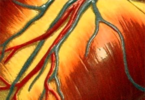

USP and MIT researchers intend to substitute damaged cardiac muscle and induce new vein formation in acute heart attack cases. This novel technique, known as cardiac reparation, uses stem cells.
USP and MIT researchers intend to substitute damaged cardiac muscle and induce new vein formation in acute heart attack cases. This novel technique, known as cardiac reparation, uses stem cells.
USP and MIT researchers intend to substitute damaged cardiac muscle and induce new vein formation in acute heart attack cases. This novel technique, known as cardiac reparation, uses stem cells.

USP and MIT researchers intend to substitute damaged cardiac muscle and induce new vein formation in acute heart attack cases. This novel technique, known as cardiac reparation, uses stem cells.
By Elton Alisson
Agência FAPESP – The treatment of ischemic heart diseases, such as acute myocardial infarction, is an area of medicine that has greatly progressed over the last few years through the development of new drugs, revascularization surgery and techniques such as angioplasty and the utilization of stents to disobtruct arteries.
A new technique promises to even further revolutionize the advances in this field. Researchers intend to substitute damaged cardiac muscles and induce the formation of new veins in patients suffering from acute myocardial infarction through the use of stem cells and embryos.
This technique, known as biological cardiac reparation, has been explored for the past ten years by several research groups, including those of José Eduardo Krieger, Director of the Molecular Genetics and Cardiology Institute (InCor) and full professor in Genetics and Molecular Medicine at the Cardiopneumology Department of the Universidade de São Paulo Medical School (FMUSP).
However, this technique must overcome diverse challenges before reaching clinical application. “Although we already have evidence that inducing the formation of new cardiac veins is possible, the much-needed substitution of damaged cardiac muscles is still a mirage,” Krieger explained during the Regional Symposium on Translational Medicine held December 2, 2011, at the FAPESP auditorium. The event was part of commemoration of the 60th anniversary of the National Scientific and Technological Development Council (CNPq).
According to Krieger, since the 1990s, there has been evidence that it is possible to induce formation of new cardiac veins and that this could be a good therapeutic target. However, the first indications that cardiac muscles have the capacity to regenerate in mammals only began to emerge in the last few years.
In a study published in 2009 in Science, a group in Sweden managed to establish the age of cardiac muscles through dating techniques that incorporated carbon 14. They examined the DNA of individuals exposed to radioactivity by nuclear bomb tests during the Cold War.
The Swedish scientists found that human cardiac muscles presented a 1% to 2% renewal capacity annually in the first decades of life and a 0.45% capacity after the fourth decade, when the probability of a myocardial infarction is greater.
The renewal rates indicated that roughly 50% of human cardiac muscle cells are substituted throughout the lifetime of individuals aged 70 years, suggesting that the development of therapeutic strategies, such as biological cardiac reparation, could stimulate this process.
“This percentage of cardiac muscle renewal in the second to fourth decades of life could be insufficient for repairing muscle damaged in a heart attack, but this very important evidence indicated that cellular renewal occurs even in this phase of life. And what we eventually want to do is explore this from a therapeutic point of view,” Krieger explained.
One of the strategies being investigated for cardiac reparation is the utilization of adult stem cells from bone marrow or adipose tissue and adult genetically modified cells to stimulate new vein formation. Nevertheless, Brazilian researchers have discovered that few cells remain in the hearts of animals when injected in the blood stream one to seven days after a myocardial cardiac infarction and that when cells are directly injected in cardiac tissues, there is a 7% increase in retention, which is still unsatisfactory.
To increase retention in heart cells, the Krieger group, in collaboration with another group at the Massachusetts Institute of Technology in the U.S., is using a series of biopolymers and natural compounds such as fibrin and collagen as a “glue” to increase fixation in heart cells.
Injected with the cells, these biopolymers, which could be extracted from the respective patient, are capable of increasing cell retention in myocardial muscles and their surrounding tissue by 15% with fibrin and up to 25% with collagen.
Krieger said, “We are learning that, in addition to making the cells remain where we want them to, these biopolymers also contribute to protecting the injected cells and facilitate the dissemination of growth factors produced for neighboring cells.”
The project, which will conclude by the end of December, was approved through a call for proposals conducted by FAPESP in collaboration with MIT.
In the laboratory at MIT, the scientists prioritized experiments with human cells. At InCor, the tests were conducted on swine cells, which are considered the closest animal model to humans.
The Brazilian team is now coordinating a study with 140 chronic ischemic patients that is in the final stages. Half of the patients submitted to bypass surgeries for myocardium revascularization will receive bone marrow cells, and the other half will receive a placebo to test whether the cells are capable of increasing blood tissue perfusion to inhibit or at least retard cardiac tissue deterioration after myocardial infarctions.
Krieger noted, “The current challenges in this area are, on the one hand, understanding the functional mechanisms by which different stem cells can contribute to minimizing cardiac damage after a heart attack and, on the other hand, evaluating whether these effects can be substituted for drugs that can be rationally investigated to evaluate the potential of this technology and its eventual application in routine clinical practice.”
Republish
The Agency FAPESP licenses news via Creative Commons (CC-BY-NC-ND) so that they can be republished free of charge and in a simple way by other digital or printed vehicles. Agência FAPESP must be credited as the source of the content being republished and the name of the reporter (if any) must be attributed. Using the HMTL button below allows compliance with these rules, detailed in Digital Republishing Policy FAPESP.


