
Nanometric modifications applied to the surface of dental implants enable researchers to accelerate the integration process between the implant and bone tissue in animal models (photo:Unesp)
Nanometric modifications applied to the surface of dental implants enable researchers to accelerate the integration process between the implant and bone tissue in animal models.
Nanometric modifications applied to the surface of dental implants enable researchers to accelerate the integration process between the implant and bone tissue in animal models.

Nanometric modifications applied to the surface of dental implants enable researchers to accelerate the integration process between the implant and bone tissue in animal models (photo:Unesp)
By Fábio de Castro
Agência FAPESP – After performing nanometric physiochemical modifications on the surfaces of dental implants, researchers from the Universidade Estadual Paulista (UNESP) were able to accelerate the osseointegration process—the union of the implant with bone tissue—in animal models.
The experiment was part of a doctoral thesis prepared by Thallita Pereira Queiroz, defended in 2010 in the UNESP Araçatuba Dental School (FOA) Oral and Maxillofacial Surgery and Traumatology Department.
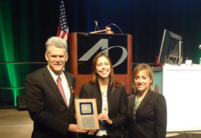
The study won Queiroz, who is now a professor at the Centro Universitário de Araraquara (UNIARA), the Best Poster Presentation Award at the North American Academy of Osseointegration’s 27th Annual Meeting held from March 1-3 2012 in Phoenix, Arizona (U.S.).
According to thesis co-advisor Antônio Carlos Guastaldi, professor at the UNESP Araraquara Chemistry Institute and responsible for implant surface development, many co-authors participated in the study due to its complexity and multi-disciplinary nature.
The result of the experiment competed, in the form of a poster, with 231 studies conducted at major universities in the United States, Europe and Asia. Only first place is awarded.
Aside from Queiroz and Guastaldi, other contributors to the work include Rogério Margonar and Ana Paula de Souza Faloni, both from the UNIARA Post-Graduate Dental Sciences Program; Professor Roberta Okamoto, study coordinator and head of the FOA-UNESP Immunohistochemical Laboratory; and Francisley Ávila Souza, Eduardo Hochuli Vieira and Idelmo Rangel Garcia Júnior, all FOA-UNESP professors.
The project received FAPESP funding through its Regular Research Support program in a project coordinated by Vieira. The implants used in the study were provided by prosthesis manufacturer Empresa Conexão Sistemas de Próteses, located in Arujá, SP.
The study aimed to promote nanometric physiochemical and morphological changes on the surfaces of dental implants to help them interact better with bone tissue. According to Guastaldi, in addition to contributing a new alternative implant surface preparation, the study changes data interpretation paradigms.
“Normally in biology, it’s thought that the morphology of the implant’s surface is more important than its chemistry in promoting osseointegration. This study inverted the paradigm, reiterating the results of our studies in recent years: the chemical modification of the surface is the most important thing for osseointegration, while morphology merely increases the area where the phenomenon occurs,” Guastaldi told Agência FAPESP.
In the study, 45 rabbits received 90 implants. After 30, 60 and 90 days, the animals were euthanized, and the implants were removed for analysis so that certain bone proteins fundamental to osseointegration could be detected.
Hydroxylapatite is the main component in tooth enamel and dentin. Changes made to the implant surfaces included modification by laser bundles, followed in some cases by hydroxylapatite displacement with biomimetics, which uses a body fluid solution with a chemical composition, temperature and pH similar to those of blood plasma.
“These implants were compared with two commercially available implants, one with its surface subjected to an acid bath and the other without any superficial modifications. All were installed in rabbit tibia for topographic, biomechanical, histometric and immunohistochemical analyses,” Guastaldi explained.
According to the professor, the study’s results showed that the nanometric physiochemical changes applied to the dental implants accelerated the expression of bone proteins that constitute the first step in the interaction between the bone and the implant.
“Bone tissue response to the implant was accelerated because of the changes, which will therefore shorten the time required for osseointegration. The next step will be to perform the same type of experiments in humans,” said Guastaldi.
Republish
The Agency FAPESP licenses news via Creative Commons (CC-BY-NC-ND) so that they can be republished free of charge and in a simple way by other digital or printed vehicles. Agência FAPESP must be credited as the source of the content being republished and the name of the reporter (if any) must be attributed. Using the HMTL button below allows compliance with these rules, detailed in Digital Republishing Policy FAPESP.


