
Study shows that vitroceramics, together with low-intensity laser light, aid in healing bone defects in rats
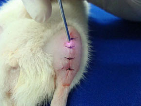
Brazilian study shows that vitroceramics, together with low-intensity laser light, aid in healing bone defects in rats.
Brazilian study shows that vitroceramics, together with low-intensity laser light, aid in healing bone defects in rats.

Study shows that vitroceramics, together with low-intensity laser light, aid in healing bone defects in rats
By Karina Toledo
Agência FAPESP – Recent studies have revealed that Biosilicato – an implantable bioactive material developed in the Vitreous Materials Laboratory (LaMaV) of the Engineering Materials Department at the Universidade Federal de São Carlos (UFSCar) – is capable of stimulating the formation of bone tissue and could be an important material for the treatment of bone fractures and dental sensitivity.
Researchers at the Santos Basin Campus of the Universidade Federal de São Paulo (Unifesp) have shown that the osteogenic effect of the material can be potentiated with by applying low-intensity laser light at the site of the implant.
The study, developed as part of the post-doctoral work of Paulo Sérgio Bossini, a FAPESP fellow, won an award at the 2013 Conference of the North American Association for Light Therapy (NAALT), held earlier this year in the United States.
Bossini began to investigate the effect of Biosilicato on the consolidation of bone defects induced in the tibias of rodents, as part of his doctoral studies under the supervision of Professor Nivaldo Antonio Parizotto in the Physiotherapy Department of UFSCar. Bossini’s post-doctoral work was conducted under the supervision of Professor Ana Cláudia Muniz Renno in Unifesp’s Biosciences Department.
“There are different presentations of Biosilicato. During my doctorate, I evaluated the usage of granules. Because it is a new biomaterial, initial tests are necessary to see if there is a risk of rejection or other adverse reactions,” explained Bossini.
Other researchers induced conditions similar to osteoporosis in rats through the extraction of their ovaries – a well-established protocol in the scientific literature. Subsequently, they created cavities in the rodents’ tibias using a drill with a diameter of approximately 3 millimeters. The objective was to simulate the defects that normally affect patients with osteoporosis.
The animals were then divided into six groups. The animals in the first group, considered the control group, only had their skin sutured and did not receive any treatment. The animals in the second group had their skin sutured and were treated with laser applications on alternate days in 60-joule doses per square centimeter (J/cm2). The animals in the third group received the same treatment in doses of J/cm2.
The fourth group was treated only with Biosilicato. The granules of the biomaterial were introduced into the defective bone with the aid of a spatula until the defect was completely covered, and the skin of each animal was then sutured. The animals in the fifth group received a 60-J/cm2 dose of laser treatment along with Biosilicato, and the animals in the sixth group received a 120-J/cm2 laser treatment along with Biosilicato.
“In analyses conducted after 14 days, the group treated with the 120 J/cm2 laser dose and Biosilicato had the largest area of newly formed bone tissue. Furthermore, in comparison to the other groups, this tissue was more mature and organized,” explained Bossini.
After completing his post-doctoral work, Bossini once again tested the combination of laser (120 J/cm2) treatment and Biosilicato – this time in a presentation known as a scaffold. The scaffold used in the study were produced during the doctoral studies of Murilo Crovacce, under the supervision of professors Ana Candida Martins Rodrigues and Oscar Peitl from DeMa-UFSCar. “The scaffold is a type of prosthetic made to the size of the defective bone but with high porosity. The objective is not to substitute for the bone, because the prosthetic has low mechanical resistance, but rather to serve as a mold and provide support for the new tissues that will grow,” explained Bossini. According to Bossini, the material is absorbed by the organism at the same time that it helps to form new bone tissue.
The same protocol was used to induce defects in rat tibia, and the animals were divided into four groups: a control group, a group treated with a laser at 120 J/cm2, a group that received only a Biosilicato scaffold, and a group that was treated with the biomaterial plus laser applications.
In the first morphological analysis, conducted 15 days after the beginning of treatment, none of the groups presented problems with rejection, necrosis or proliferation of atypical cells.
The group that received only laser applications had the largest area of newly formed bone. According to Bossini, this is due to the laser’s ability to stimulate the formation of new blood vessels at the site and to recruit blood cells.
“Although the Biosilicato stimulates the formation of bone tissue, it also needs time to be degraded by the organism. In the first moment, the scaffold could work as a physical barrier to tissue growth, but it is already perceptible that the prosthetic shrinks, allowing room for the newly formed bone,” said Bossini.
Immunohistochemical analysis (which evaluates the proteins found in the tissue), by contrast, revealed that the group that received Biosilicato plus laser treatments had greater expression of factors related to bone tissue formation – especially RUX-2 and COX-2 proteins – in the cells surrounding the site of the implant.
“This is clear evidence that the material has osteogenic potential and that this effect was potentiated with laser applications,” affirmed Bossini.
The researchers are currently analyzing data from the examinations conducted 30, 45 and 60 days after the beginning of treatment to determine whether the biomaterial has been completely absorbed by the rats and whether it favors the formation of better-organized tissue. The preliminary data indicate that the newly formed bone has the same biomechanical characteristics as healthy bone.
Furthermore, the scientists are analyzing gene expression in the newly formed bone tissue area, and the preliminary results reveal that the biomaterial induces higher expression of genes associated with bone repair, explained Bossini.
Several presentations
Biosilicato is a bio-glass developed in the 1990s at LaMaC under the supervision of the UFSCar Professor Edgar Dutra Zanotto and patented in 2004.
The material is consists of sodium, potassium, calcium, phosphorus, oxygen and silicon. It can be implanted at the treatment site in the form of granules, scaffolds, or fibers or as a single (monolithic) piece, made to size to replace, for example, one of the bones in the ear.
“The material is partially soluble in blood plasma and connects to bone, teeth or cartilage. A layer of hydroxycarbonate apatite, a substance that has the same composition as the mineral tissue of teeth and bones, forms on its surface,” affirmed Zanotto.
“In addition to the remineralizing potential, recent studies have shown that Biosilicato accelerates the formation of bone cells, but this mechanism still needs to be better studied,” explained Zanotto, who also coordinates the Glass Research, Education and Innovation Center (CEPIV), one of the new Research, Innovation and Dissemination Centers (CEPID).
Republish
The Agency FAPESP licenses news via Creative Commons (CC-BY-NC-ND) so that they can be republished free of charge and in a simple way by other digital or printed vehicles. Agência FAPESP must be credited as the source of the content being republished and the name of the reporter (if any) must be attributed. Using the HMTL button below allows compliance with these rules, detailed in Digital Republishing Policy FAPESP.


