


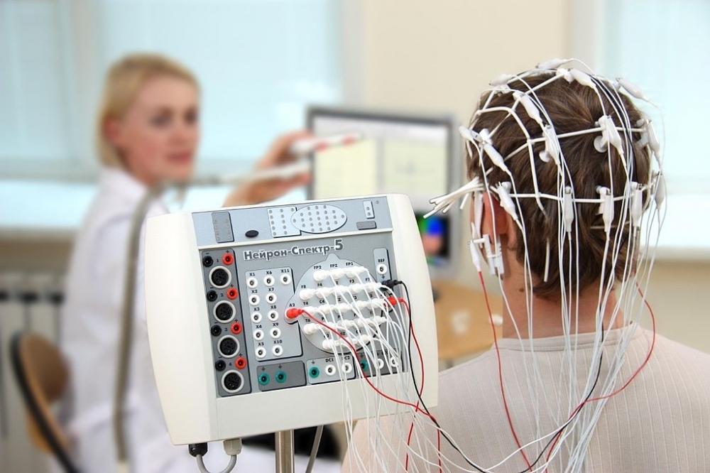
In a lecture given at FAPESP Week France, Brazilian scientist Francisco Fraga da Silva spoke about developing a low-cost diagnostic alternative based on electroencephalography data (photo: Baburov / Wikimedia Commons)
In a lecture given at FAPESP Week France, Brazilian scientist Francisco Fraga da Silva spoke about developing a low-cost diagnostic alternative based on electroencephalography data.
In a lecture given at FAPESP Week France, Brazilian scientist Francisco Fraga da Silva spoke about developing a low-cost diagnostic alternative based on electroencephalography data.
In a lecture given at FAPESP Week France, Brazilian scientist Francisco Fraga da Silva spoke about developing a low-cost diagnostic alternative based on electroencephalography data (photo: Baburov / Wikimedia Commons)
By Heitor Shimizu, from Paris | Agência FAPESP – With the aim of enabling the early diagnosis of Alzheimer’s disease, researchers at the Federal University of the ABC – UFABC in Greater São Paulo, Brazil are working to develop, improve, and validate markers based on a computer analysis of quantitative electroencephalography data, which involves a low-cost and non-invasive technique that enables brain activity to be mapped.
The work is coordinated by Francisco José Fraga da Silva, a professor at UFABC’s Center for Engineering, Modeling, and Applied Social Sciences (CECS), who spoke about the topic in a lecture given at FAPESP Week France.
“Although it’s a progressive disease and, up to now, incurable, early diagnosis and treatment enable the progression of symptoms to be delayed and an improvement in patients’ quality of life,” said the researcher.
During the event, Fraga da Silva presented a summary of the studies that he has conducted in the last ten years – various ones with FAPESP’s support – on processing and classifying quantitative electroencephalography tests. The aim is to create methods to support the early diagnosis of both Alzheimer’s disease and a condition known as mild cognitive impairment (MCI), characterized by mild and continuous memory loss, which may or may not develop into dementia.
The research involves information engineering, neuroscience, and topics such as biomedical signal processing, pattern recognition, and machine learning.
“It’s very important to diagnose and monitor patients with MCI appropriately, as they’re people with an increased risk of developing dementia,” said Fraga da Silva. “Recent statistics show that around 50% of all people who reported symptoms of MCI to a doctor developed Alzheimer’s within four years.”
The researcher explained that there are already validated methodologies for diagnosing MCI and Alzheimer’s that require equipment such as CT and fMRI scanners – both of which are expensive and difficult to access in poorer countries or in areas far from large urban centers.
The electroencephalogram, on the other hand, is a cheap, portable, and, moreover, non-invasive technique. The equipment can record the brain’s electrical activity via electrodes placed on the scalp.
According to Fraga da Silva’s assessment, the low cost and widespread availability of the equipment enable serial exams to be carried out and the monitoring of the evolution of neurological states.
Results
In a study recently published in the journal Computer Methods and Programs in Biomedicine, the group which comprises UFABC and Canadian collaborators investigated the use of event-related potentials (ERP) and event-related synchronization/desynchronization (ERS/ERD) to diagnose early-stage Alzheimer’s disease.
ERP is a type of analysis based on the average of various electroencephalogram segments that enables specific brain activity to be identified when the individual is exposed to certain sensory stimuli (generally visual or auditory).
ERS/ERD analysis allows for increases and decreases to be detected, respectively, in specific rhythms (alpha rhythm, for example) that occur during or shortly after sensory stimulation.
The electroencephalogram covers four main brainwave rhythms: beta (normal waking state), alpha (relaxed mental states), theta (occurs during meditation, sleep, and deep relaxation), and delta (deep sleep).
“These rhythms, which are nothing more than oscillations in specific frequency bands, usually appear combined in electroencephalogram signals, but they can be separated using filters,” Fraga da Silva explained to Agência FAPESP.
The authors of the article compared – during the realization of tasks that required the use of short-term memory (working or operational memory) – the behavioral results (reaction time and accuracy) and the ERP and ERS/ERD responses in healthy elderly people (control group), in patients with MCI, and in another group of early-stage Alzheimer’s sufferers.
The results indicated that the ERS/ERD analyses were more efficient than ERP in the early detection of Alzheimer’s and MCI.
“We hope that from the search for topographic biomarkers based on quantitative electroencephalography a computer app can emerge that enables the accuracy, sensitivity, and specificity in the early detection of Alzheimer’s disease to be significantly increased,” said Fraga da Silva.
The FAPESP Week France symposium was held between November 21st and 27th, thanks to a partnership between FAPESP and the universities of Lyon and Paris, both in France. Read other news about the event at http://www.fapesp.br/week2019/france/.
The Agency FAPESP licenses news via Creative Commons (CC-BY-NC-ND) so that they can be republished free of charge and in a simple way by other digital or printed vehicles. Agência FAPESP must be credited as the source of the content being republished and the name of the reporter (if any) must be attributed. Using the HMTL button below allows compliance with these rules, detailed in Digital Republishing Policy FAPESP.
