


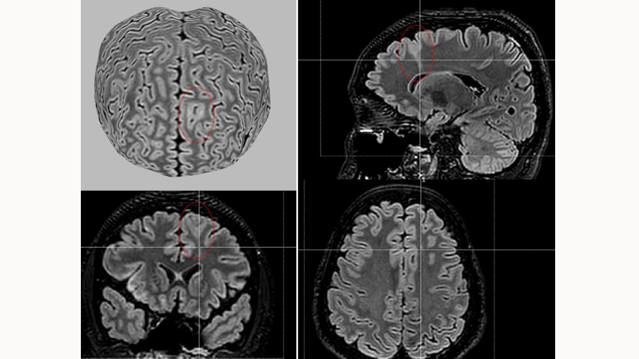
Magnetic resonance image reconstruction showing a subtle lesion of focal cortical dysplasia (indicated by the red circle) in a child with drug-refractory epilepsy (credit: Laboratory of Neuroimaging/BRAINN/UNICAMP)
Publicly available algorithm facilitates lesion identification and surgical planning for patients with focal cortical dysplasia, a malformation associated with a drug-refractory form of the disease.
Publicly available algorithm facilitates lesion identification and surgical planning for patients with focal cortical dysplasia, a malformation associated with a drug-refractory form of the disease.
Magnetic resonance image reconstruction showing a subtle lesion of focal cortical dysplasia (indicated by the red circle) in a child with drug-refractory epilepsy (credit: Laboratory of Neuroimaging/BRAINN/UNICAMP)
By Maria Fernanda Ziegler | Agência FAPESP – An international team of researchers has developed a tool based on artificial intelligence (AI) that identifies brain lesions associated with focal cortical dysplasia, one of the main causes of drug-refractory epilepsy and very difficult to diagnose. The details were published in the journal JAMA Neurology.
Focal cortical dysplasia is a malformation of the cortex characterized by lesions in certain regions of the brain. Patients with the condition tend to be resistant to treatment with anticonvulsants, leaving surgery (to remove the damaged areas) as the only alternative to reduce the symptoms of the disease. However, these brain lesions are often very subtle and difficult to detect, making the treatment of focal cortical dysplasia even more challenging.
Using machine learning techniques and analyzing MRI data from 1,185 participants – including 703 people with focal cortical dysplasia and 482 controls – the tool, called MELD Graph, was able to detect the abnormalities from scans of the brain images.
The tool, whose algorithm is publicly available, detected 64% of brain abnormalities associated with epilepsy that radiologists had missed. As a result, MELD Graph is expected to be able to help more than 4 million people worldwide living with this cause of epilepsy.
International collaboration
The work was done as part of the Enigma Consortium, an international network that brings together scientists from fields including imaging genomics, neurology and psychiatry. The group seeks to understand the structure and function of the brain using high-resolution magnetic resonance imaging, genetic data and other information from patients with epilepsy, Parkinson’s, Alzheimer’s, autism, schizophrenia and other neurodegenerative diseases.
The AI tool was developed by researchers at King’s College London and University College London, both in the United Kingdom. The high-quality magnetic resonance images were provided by 23 epilepsy research centers around the world that are part of the consortium. The Brazilian Institute of Neuroscience and Neurotechnology (BRAINN) was one of the institutions with a high image acquisition and analysis capacity to provide data for the project.
“In this type of epilepsy, patients – who can be adults or children – have 30 or even 50 epileptic seizures a day. It’s serious, and usually the seizures aren’t controlled by medication. Therefore, surgery to remove the brain lesions, although very difficult to perform, is the best option for these cases. The new tool can greatly help these patients, speeding up the treatment and, above all, improving the quality of life of these people and their families,” Clarissa Yasuda, professor at the Department of Neurology of the School of Medical Sciences of the State University of Campinas (FCM-UNICAMP) and member of BRAINN, a FAPESP Research, Innovation and Dissemination Center (RIDC), told Agência FAPESP.
According to Luca Palma of the Bambino Gesù Children’s Hospital in Italy, the MELD Graph identified a subtle lesion that many radiologists missed in a 12-year-old boy who had daily epileptic seizures and had tried nine anticonvulsant drugs without improvement. “This tool can identify epilepsy patients who can be treated surgically and help plan surgery – reducing risks, saving money and improving outcomes,” the researcher said in a press release.
In addition to reducing costs and improving patients’ quality of life, another important benefit of the new tool is the opportunity to expand our understanding of focal cortical dysplasia. “It’s a brain malformation about which there’s still much to be learned. By analyzing these high-quality images and using the tool, we’ll be able to identify patterns that would be impossible to see without artificial intelligence and answer questions such as the origin of this problem,” says Fernando Cendes, professor at FCM-UNICAMP and BRAINN coordinator.
The article “Detection of Epileptogenic Focal Cortical Dysplasia Using Graph Neural Networks A MELD Study” can be read at: jamanetwork.com/journals/jamaneurology/fullarticle/2830410?guestAccessKey=e833d51e-69c6-4a26-bee3-66b84e3e9b0c&utm.
The Agency FAPESP licenses news via Creative Commons (CC-BY-NC-ND) so that they can be republished free of charge and in a simple way by other digital or printed vehicles. Agência FAPESP must be credited as the source of the content being republished and the name of the reporter (if any) must be attributed. Using the HMTL button below allows compliance with these rules, detailed in Digital Republishing Policy FAPESP.
