


Brazilian researchers analyze the structure of the neurons in Paint Horse intestines using three-dimensional resource
Brazilian researchers analyze the structure of the neurons in Paint Horse intestines using three-dimensional resource.
Brazilian researchers analyze the structure of the neurons in Paint Horse intestines using three-dimensional resource.
Brazilian researchers analyze the structure of the neurons in Paint Horse intestines using three-dimensional resource
By Noêmia Lopes
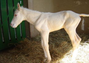
Agência FAPESP – Ileocolonic aganglionosis is a genetic disease that affects the peristaltic movements and absorption of nutrients in the intestines of the Paint Horse, an expensive breed of horse favored by animal owners. The disease is fatal and kills 100% of animals in the first few years of life. Hirschsprung disease is a similar illness found in humans that affects one in 5,000 children in a milder, more treatable way.
The origin of the two problems is a type of deficiency or lack of migration in the cells from the neural crest – cells in the central nervous system that travel throughout the body of embryos to form the peripheral nervous system, including the intestines (which include the enteric nervous system).
In an article published by Cells Tissues Organs on July 25, Universidade de São Paulo (USP) and Universidade Estadual Paulista (Unesp – Botucatu Campus) researchers detailed their three-dimensional analysis of the enteric neurons of horses. The neurons were obtained from the ileum (the part of the intestine that is most affected by aganglionosis in equines).
“The analysis of enteric horse neurons through 3-D stereology – science that, in our case, refers to the study and quantification of biological particles, neurons in three dimensions – is an important achievement for neuroscience. We did not find anything similar in the national and international literature,” said Professor Antonio Augusto Coppi, research coordinator responsible for the Laboratory of Stochastic Stereology and Chemical Anatomy (LSSCA) of USP’s Veterinary Medicine and Zootechny Department.
Until recently, the enteric neurons were studied only through bidimensional methods based on the shape defined by the area and the diameter of these cells, which resulted in imprecise estimates of the total number and size.
“The technique of 3-D quantitative microscopy used by stereology allows more accurate calculations of the total number, the surface and the real size of cells. With this information, we can better understand the structural effects of the disease on certain cells, which can facilitate possible future genetic treatments for horses and advances in the therapeutic schemes used to treat Hirschsprung disease in humans,” explains Coppi.
Ten colts – five healthy and five with ileocolonic aganglionosis – underwent blood exams (to genetically confirm the presence of the disease) and clinical exams (to evaluate whether they managed to defecate while prostrate or active, among other parameters). After seven days, the animals were euthanized according to existing legislation, and researchers removed their ileum.
Utilizing specific 3D software installed in a microscope adapted to record the position of cells on the Z axis (or rather, by depth in the tissue), the team obtained a 3-D spatial sample of the neurons found in the ileum of the animals. This procedure involved measuring the length, width and depth, in addition to estimates of volume, surface and total number. “It’s like looking at the ocean from the top to the bottom and observing schools of fish of varied sizes and shapes at different depths,” compares Coppi.
Although it makes the process more rapid, the high-cost software is not necessary. According to the researcher, “3-D stereology can be performed by generating sequential physical planes. Or, rather, working first with areas of plane A or with those of plane B, estimating the distance between the two (generating depth) and, therefore, obtaining volumes.”
According to Coppi, certain researchers work with quantitative bidimensional microscopy because they believe that the neurons in the intestine are arranged in a single layer of cells. His impression is different, as he stated that enteric neurons are organized in more than one layer. “We found two to three layers of enteric neurons in the current study,” he affirms.
Quantity and size of the neurons
In the gastrointestinal tube, there are two types of nerve cells. The myenteric neurons are distributed in the muscular layer of the ileum and coordinate peristaltic movements throughout the length of this tube. However, submucous neurons located in the submucosal layer of the organ are responsible for controlling gastrointestinal secretion, the local blood flow and the absorption of nutrients.
In the ileum of the sick animals, the researchers observed that the number of myenteric neurons was reduced by 99%. Because it was not a complete reduction, they proposed a change in nomenclature to hypoganglionosis (the hypo refers to a diminished number and not to a total lack of neurons) for this specific disease profile. The remaining neurons (1%) were approximately one-half of their normal size (63.8%). This condition indicates severe atrophy and makes it impossible for the neurons to function properly.
The drastic reduction in the myenteric neurons and the atrophy of the remaining cells initiate a week of diarrhea and dehydration, followed by the interruption of peristaltic movement and intestinal function. Toxins accumulate and enter the circulatory system, and the animal dies. Medicine is ineffective because it works by stimulating these neurons, which are practically nonexistent or inoperative in cases such as those examined in the study.
The submucous neurons were completely absent in the ileum of the sick horses. This condition makes it difficult to absorb the colostrum of the mother and causes the colt to become malnourished, which also contributes to the death of the animal.
“Thanks to the 3D stereology technology, we know the spatial structure of the intestinal nervous cells in detail and know how they are organized within the organ. We believe that in the future, these data can facilitate genetic therapy for treating the disease in horses and can create new ways to approach Hirschsprung in humans,” affirms Coppi.
The results were obtained through a FAPESP research grant and a fellowship.
“We will continue this project because, in addition to the ileum, we extracted other portions of the intestines of the foal, which are still being analyzed,” adds Coppi. Other participants include doctoral student Aliny Ladd and Fernando Ladd (PhD) and Andrea Almeida (PhD) of LSSCA/USP; Professors Carla Belli, Luis Cláudio da Silva and André de Zoppa of FMVZ/USP; and Professor Alexandre Borges, of Unesp – Botucatu’s Veterinary Medicine and Zootechny School (FMVZ).
More information is available at: www.lssca.fmvz.usp.br, guto@usp.br and LSSCA’s Facebook page.
The Agency FAPESP licenses news via Creative Commons (CC-BY-NC-ND) so that they can be republished free of charge and in a simple way by other digital or printed vehicles. Agência FAPESP must be credited as the source of the content being republished and the name of the reporter (if any) must be attributed. Using the HMTL button below allows compliance with these rules, detailed in Digital Republishing Policy FAPESP.
