


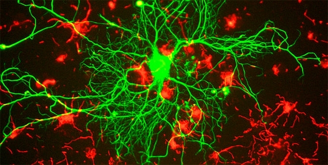
Study conducted in Brazil aims to identify alterations to motor neurons in the presymptomatic stage of ALS (image: Gerry Shaw / Wikipedia)
Study conducted in Brazil aims to identify alterations to motor neurons in the presymptomatic stage of ALS.
Study conducted in Brazil aims to identify alterations to motor neurons in the presymptomatic stage of ALS.
Study conducted in Brazil aims to identify alterations to motor neurons in the presymptomatic stage of ALS (image: Gerry Shaw / Wikipedia)
By Karina Toledo | Agência FAPESP – Researchers at the University of Campinas (UNICAMP) in São Paulo State, Brazil, have developed a mathematical model for computer simulation of the alterations that occur in the motor neurons of people with amyotrophic lateral sclerosis (ALS), the disease that afflicted British physicist Stephen Hawking.
Characterized by progressive muscular paralysis, ALS is caused by genetic mutations that impair the production or activity of SOD1 (copper-zinc superoxide dismutase-1), an enzyme that keeps nerve cells safe from toxic metabolic waste. These mutations may or may not be inherited. The condition leads to degeneration and death of the motor neurons responsible for innervating skeletal muscles and controlling voluntary movement.
The main aim of the group led by Professor Leonardo Abdala, who heads the Neuroengineering Research Laboratory in UNICAMP’s School of Electrical Engineering & Computation, is to understand the molecular mechanisms associated with neuron degeneration, including alterations in the functioning of proteins permeable to calcium, sodium and potassium ions (ion channels) that may affect neuron response and hence the control of muscle power.
“We used a computer simulation to look for a biological marker in the presymptomatic stage of the disease – a biophysical phenomenon that occurs in motor neuron membranes and affects the cell’s electrical activity and control of muscular force. This would pave the way for the study of pharmacological interventions capable of reversing or mitigating the problem,” said Elias, who also heads UNICAMP’s Biomedical Engineering Center.
Mammals have two types of motor neurons, upper and lower, Elias explained. Upper motor neurons originate in the motor cortex of the brain and send electrical impulses to the lower motor neurons, which are located in the spinal cord and brain stem. These nerve impulses are relayed to the muscles, which convert them into movements.
During the master’s research of Débora Elisa da Costa Matoso, the group developed a mathematical model to simulate the dynamics of a lower motor neuron both in a healthy state and in a condition equivalent to ALS. The model was based on data from experiments with rodents published in the scientific literature.
“No data for human patients is available, only for animal models in mice that are genetically modified to reproduce a condition resembling ALS,” Elias explained. “To validate the model, we simulated the experiments performed with animals in the lab. The results were compatible with those obtained in vivo and in vitro. This suggests that the model can indeed represent what happens to lower motor neurons during progression of the disease in mice.”

Motor neuron: left, representation of the biological system; center, representation of the mathematical model; right, representation of the computational model (illustration: Débora Matoso)
The group uses computer simulations mainly to study what happens in three ion channels: one permeable to sodium ions, normally located in the neuron’s cell body; another permeable to calcium, normally located in its dendritic branches; and a third channel permeable to potassium, found both in the cell body and in the dendrite.
According to Elias, the results thus far suggest that the potassium channel is fundamental to explaining some important alterations observed in the dynamics of the lower motor neuron, although only scarce data from experiments with animals are available to corroborate this finding.
“The only drug currently available to treat ALS, riluzole, acts on the persistent sodium channels. If we can show with the model that other ion channels are also involved in this process of degeneration, we can make room for new research to be done in animals to test new pharmacological interventions,” Elias said.
Matoso now intends to develop a complete model of the neuromuscular system during her PhD research to investigate how the biophysical mechanisms that alter motor neuron dynamics influence the production of force in the initial stage of the disease.
“In parallel, we plan to perform experiments in partnership with groups that have access to ALS patients to try to collect as much data as possible to validate the model we’re developing,” Elias said. “As a second step in this research, we’ll compare the results of the simulations with the experimental results in search of potential clinical and pharmacological interventions.”
In addition, Elias has established a partnership with a team from the Brazilian Research Institute for Neuroscience and Neurotechnology (BRAINN) led by Professor Li Li Min with the aim of studying the effects of different neuromodulation techniques (application of electrical current, transcranial magnetic stimulation, and focused ultrasound) on force control in stroke patients and individuals with Parkinson’s disease or cerebellar ataxias (a group of diseases that affect movement control).
BRAINN Congress
Elias presented the results of his research on April 10, 2018, in Campinas during the 5th BRAINN Congress, held by BRAINN, one of the Research, Innovation and Dissemination Centers (RIDCs) funded by FAPESP.
Among the event’s international highlights was a presentation by Professor John A. Detre, a researcher at the University of Pennsylvania in the United States, on the use of functional neuroimaging technologies such as magnetic resonance imaging (MRI) and optical imaging to study brain function in healthy individuals and in patients with a variety of clinical disorders including stroke, epilepsy, neurodegenerative diseases and migraine.
“In an adult individual, the brain uses approximately 20% of blood flow even though it corresponds to only 2% of the body’s mass. Because blood flow and metabolism are closely coupled, we can use flow measurements to study many aspects of brain function. To do so, we use functional imaging techniques,” Detre told Agência FAPESP.
Among the Brazilian highlights of the conference program was Roberto Lent, director of the Federal University of Rio de Janeiro’s Biomedical Science Institute (ICB-UFRJ). Lent’s presentation focused on studies of neuroplasticity, the ability of the nervous system to change structurally and functionally during neuronal development and in response to environmental interference.
“Environmental interference may be a disease, a conversation between people, or an educational action,” Lent said. “When a professor teaches something, she changes the student’s brain, and this is one kind of plasticity. In my lab, we address long-distance plasticity, which is the capacity for change and alteration in the most important cerebral pathways.”
As a study model, the ICB-UFRJ group uses an important communication pathway between the left and right hemispheres called the corpus callosum, which comprises 200 million nerve fibers.
“The corpus callosum changes in various situations, such as after the traumatic amputation of a limb,” Lent said. “In these cases, many people develop phantom limb syndrome. They feel pain, itching, and anomalous positions of the missing arm or leg. This is the result of reorganization of the brain, especially the broad avenue that is the corpus callosum.”
The conference program also featured a roundtable on “BRAIN(N): past, present and future”, which discussed ways of boosting the impact of neuroscience research performed in Brazil and optimizing the resources allocated to it. Other participants in addition to Lent and Detre included Fernando Cendes, BRAINN’s principal investigator, Canadian researcher Richard Frayne (University of Calgary), and FAPESP Scientific Director Carlos Henrique de Brito Cruz.
The Agency FAPESP licenses news via Creative Commons (CC-BY-NC-ND) so that they can be republished free of charge and in a simple way by other digital or printed vehicles. Agência FAPESP must be credited as the source of the content being republished and the name of the reporter (if any) must be attributed. Using the HMTL button below allows compliance with these rules, detailed in Digital Republishing Policy FAPESP.
