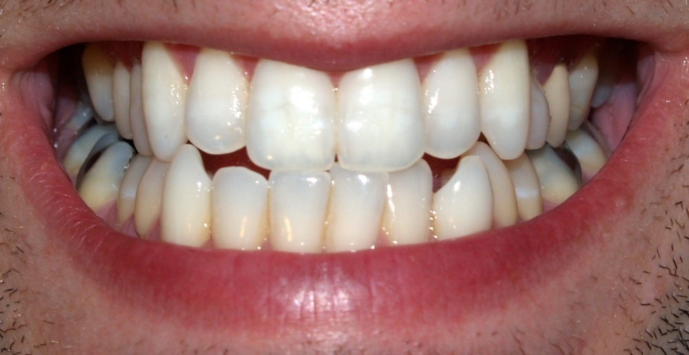

Developed by Brazilian researchers, computer model can improve evaluation of premature contact, which occurs when a restoration is larger than the original tooth (photo: David Shankbone / Wikimedia Commons)
Developed by Brazilian researchers, computer model can improve evaluation of premature contact, which occurs when a restoration is larger than the original tooth.
Developed by Brazilian researchers, computer model can improve evaluation of premature contact, which occurs when a restoration is larger than the original tooth.

Developed by Brazilian researchers, computer model can improve evaluation of premature contact, which occurs when a restoration is larger than the original tooth (photo: David Shankbone / Wikimedia Commons)
By Maria Fernanda Ziegler | Agência FAPESP – Premature contact, which is observed fairly often in patients undergoing dental surgeries and which can lead to bone loss, occurs when a filling, crown or root canal restoration is higher than the original tooth so that it and the opposite tooth are the first to touch, resulting in overloading.
Although the problem is recurrent and easily perceived by the patient, who should tell the dentist about it and have it corrected, no studies in the scientific literature have fully characterized the mechanisms relating to the bone alterations caused by microscopic changes in bone tissue tension and deformation – until now.
Researchers at the University of Campinas’s Piracicaba Dental School (FOP-UNICAMP) in São Paulo State, Brazil, have developed a computer model that simulates the action of premature contact and can be used for a better understanding of losses to the alveolar bone that supports the teeth. The innovation can also be used to study other dental problems.
The study, which was supported by FAPESP via the project “Bone remodeling and biomechanical analysis of maxillary alveolar bone in rats with masticatory overload”, initially showed (on the basis of experiments with rats) that premature contact correlates with expression of proteins in the region where bone loss occurs.
“We succeeded in observing the entire mechanism triggered by the altered mechanical stimulus,” said Ana Claudia Rossi, one of the lead investigators for FOP-UNICAMP’s Mechanobiology Research Laboratory. “When premature contact occurs, the protein RANK-L is expressed more intensely, and this causes alveolar bone loss. In parallel with the experiment in rats, we developed a computer model to simulate this cause-and-effect relationship between the protein and bone loss.”
In the study, the researchers analyzed 50 two-month-old male rats of the species Rattus norvegicus albinus. Premature contact was induced in 40 rats by cementing a 1 mm metal wire to the upper right-hand first molar.
The animals were divided into four groups according to sacrifice period (7, 14, 21 and 28 days after application of the wire), and a fifth group without any occlusal alteration served as control.
“Bone loss occurred in the first week,” Rossi said. “Microscopic analysis showed alveolar bone microdeformation at the point where the protein was expressed, indicating that the premature contact induced in the rats acted as an ‘altered’ mechanical stimulus, resulting in the initial periods in compressive microdeformation in bone tissue supporting the tooth.”
Bone deformation was quantified by computer simulation using a virtual model of the rat skull and jaws. Protein expression was analyzed by means of an immunohistochemical study relating to osteoclast resorption of RANK-L. Osteoclasts are bone cells that absorb bone tissue during growth and healing.
“The computer model enabled us to produce a mechanical and biological map of the changes that occur in alveolar bone in response to the simulated biomechanical alterations caused by premature contact,” Rossi said.
A better understanding of occlusal alterations was afforded by the results of the experiments and computer simulations combined. “We believe this will pave the way for the establishment of knowledge for clinical applications in dentistry,” she went on. “The tool can contribute to an understanding of alveolar bone physiological, dynamic and biomechanical processes under these conditions. Another interesting aspect of our research is that the virtual model, which closely approximates reality, can be used for other dental studies and so will reduce the use of animals in experiments.”
More research is required to increase the precision of the computer simulation. “Future analysis can be performed individually by simulating the condition of each patient so that both problems and solutions in terms of treatment can be better adapted to the patient’s biology and mechanics,” Rossi said.
The results were presented at the 34th Annual Meeting of the Brazilian Society for Dental Research (SBPqO), the Brazilian chapter of the International Association for Dental Research (IADR), and was awarded first prize for oral presentations on basic research.
Republish
The Agency FAPESP licenses news via Creative Commons (CC-BY-NC-ND) so that they can be republished free of charge and in a simple way by other digital or printed vehicles. Agência FAPESP must be credited as the source of the content being republished and the name of the reporter (if any) must be attributed. Using the HMTL button below allows compliance with these rules, detailed in Digital Republishing Policy FAPESP.


