


A study published in Science shows that cells transfer many more mitochondria than necessary to other cells, even at the expense of their own lives
A study published in Science shows that cells transfer many more mitochondria than necessary to other cells, even at the expense of their own lives.
A study published in Science shows that cells transfer many more mitochondria than necessary to other cells, even at the expense of their own lives.
A study published in Science shows that cells transfer many more mitochondria than necessary to other cells, even at the expense of their own lives
By Elton Alisson
Agência FAPESP – In nature, there are several species (in addition to humans) that make altruistic gestures to guarantee the survival of their offspring. Some extreme examples are female polar bears, which gain as much as 200 kilograms during gestation and fast for the first 8 months of the their cubs’ lives to provide milk rich in fat, and “mother” spiders of the Stegodyphus species, which allow their offspring to kill them so that they can serve as a food source.
A study published in Science magazine conducted by an international group of researchers, which included two Brazilians, revealed that even the smallest parts of living material – cells – also make sacrifices to guarantee the continuity of future generations.
The researchers found that during the process of cellular division (mitosis), by which a “mother” cell divides to create a “daughter” cell, the maternal cell provides many more mitochondria (internal structures essential to the survival of any cell) to its offspring than would be expected given the ratio between the two volumes (the daughter cell is smaller than the mother cell).
This discovery suggests that mother cells sacrifice themselves to increase the probability of their daughters’ survival, much like other reproductive examples found in nature.
“This realization is unprecedented and contradicts intuition that the mitochondria divide proportionally to the density [ volume] of mother and daughter cells. They break this rule,” commented one of the authors of the study, Luciano da Fontoura Costa, professor at the Physics and Informatics Departments at the University of São Paulo’s Physics Institute at São Carlos (USP São Carlos).
Costa, one of the main researchers involved in a Thematic Project conducted with FAPESP funding and coordinated by Professor Roberto Marcondes Cesar, Jr. (Department of Computer Science at USP’s Institute of Mathematics and Statistics), is working on a research project with York University in the United Kingdom under an FAPESP cooperation agreement.
The researcher served as a mentor for FAPESP Fellow Matheus Palhares Viana, the second researcher in the study, during the latter’s doctoral and post-doctoral studies.
Viana is currently pursuing post-doctoral studies at the University of California at Irvine in a research group coordinated by Susanne Rafelski – the first author of the study – with Professor Wallace Marshall of the University of California at San Francisco. Researchers from the University of Beijing also participated in the study.
To study the process of mitochondria transfer between cells, the researchers used Saccharomyces cerevisiae yeast, which is commonly used to produce bread and beer.
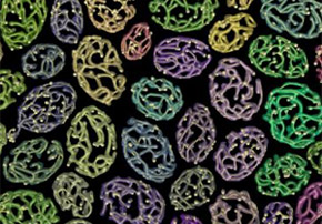
Using sophisticated microscopy techniques, the international team – lead by Marshall and Rafelski – captured images of yeast cells in a confocal fluorescence microscope and proceeded to optically slice mitochondria found in the cells. Optical slicing is a technique through which structures are fragmented into several pieces, like pieces of a puzzle.
By employing special computing methods for image processing – initially developed as part of other Thematic Projects, conducted with FAPESP funding, in which Costa also participated – the researcher and Viana put the “slices” of the mitochondria together, creating a three-dimensional reconstruction of the structures, and presented them in the form of networks (graphs). In this manner, they managed to provide detailed reproductions and measure the physical size of the mitochondrial networks, which tends to decrease over successive generations of the cells.
The researchers observed that in the case of yeast cells, however, the size of the mitochondrial network increases with the growth of the cells, scaling with the square root of the cells’ size.
“If the mitochondria were randomly divided and the density of cells was constant, we would expect to find fewer mitochondria in daughter cells than mother cells. What we discovered in this project is that the mother cell gives more mitochondria than expected to the daughter cell,” said Costa in an interview with Agência FAPESP.
According to the researchers, instead of the mother yeast supplying enough mitochondria to descendants to guarantee survival, they transfer many more organelles than are necessary, even at the cost of their lives. In the study, the majority of the cells began to die after 10 generations.
Mutant forms of the yeast, much more than “miserly” in supplying their mitochondria to future generations, lived for much longer periods.
Complementary approach
According to Costa, the discovery of these division mechanisms could be extended to other organisms and tissues. Human stem cells and some cancerous cells, for example, often divide into two cells that appear to behave very differently.
Thus, in the opinion of the researchers, biological studies of similar systems that involve the use of exact scientific approaches, such as the quantitative methods of mathematics, physics and computing and molecular analysis, complement genetics research.
According to Costa, genomic studies—today, conducted on lower scales than those of molecular biology studies—are insufficient in understanding the organism as a whole because, for example, several genes are not expressed.
“Genes, in principle, indicate how to construct a protein, for example. But having a gene does not mean the organism will have this given protein expression,” said Costa.
“There is a control [system] in the cellular machinery that determines if this protein will be expressed or not. And this control, depends on the geometry of the embryo and whether certain tissues and anatomical structures have been formed that are used as signals for gene expression and serve as platforms to build the rest of the organism.”
The article “Mitochondrial network size scaling in budding yeast” (doi:10.1126/science.1225720) by Luciano da Fontoura Costa et al. can be accessed at www.sciencemag.org/content/338/6108/822.full.
The Agency FAPESP licenses news via Creative Commons (CC-BY-NC-ND) so that they can be republished free of charge and in a simple way by other digital or printed vehicles. Agência FAPESP must be credited as the source of the content being republished and the name of the reporter (if any) must be attributed. Using the HMTL button below allows compliance with these rules, detailed in Digital Republishing Policy FAPESP.
