

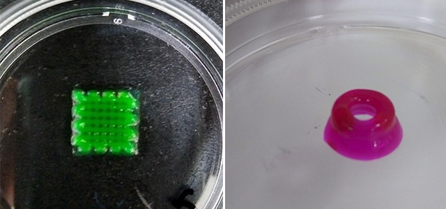
The technique can contribute to a deeper understanding of neurodegenerative diseases and lead to applications in drug testing. In the future, this technique could be used to reconstruct damaged parts of the brain (image: Bruna Alice Gomes de Melo)
The technique can contribute to a deeper understanding of neurodegenerative diseases and lead to applications in drug testing. In the future, this technique could be used to reconstruct damaged parts of the brain.
The technique can contribute to a deeper understanding of neurodegenerative diseases and lead to applications in drug testing. In the future, this technique could be used to reconstruct damaged parts of the brain.
The technique can contribute to a deeper understanding of neurodegenerative diseases and lead to applications in drug testing. In the future, this technique could be used to reconstruct damaged parts of the brain (image: Bruna Alice Gomes de Melo)
By André Julião | Agência FAPESP – Researchers at the Federal University of São Paulo (UNIFESP) in Brazil are developing a bioink capable of producing 3D neural tissue to simulate the human brain. This technology could permit a deeper investigation of neurodegenerative disorders, such as Parkinson’s disease and Alzheimer’s disease.
The idea is to reproduce the functions of the central nervous system more accurately than what can be done with traditional methods involving two-dimensional culture of a single cell type in the laboratory or studies performed in mice, which have a genome comparable to ours but a much less complex brain.
A 3D bioprinter will be used to deposit many layers of the bioink until a structure similar to a tissue or organ is built. This technology has been tested by several research groups around the world. Scientists expect it to be used to make biomaterials for transplantation in the future.
Miniature organs can now be bioprinted for use as experimental models to test drugs and study disease mechanisms. Tests performed by researchers in Brazil and elsewhere have shown brain cells to be the hardest to bioprint because of the complexity of the central nervous system, which is made up of different cell types that interact in poorly understood ways.
“The goal of this study is to develop a 3D model, more complex and closer to an in vivo model, in which we can explore cellular mechanisms of neurodegenerative diseases,” said Bruna Alice Gomes de Melo, a postdoctoral researcher who is carrying out an internship at UNIFESP’s Medical School (EPM) with a scholarship from FAPESP.
Melo presented the study at the 34th Annual Meeting of the Federation of Brazilian Societies for Experimental Biology (FeSBE), held in September 2019 in Campos do Jordão, São Paulo State. The study was supported by FAPESP as part of a recently approved Thematic Project.
“Bioprinting is at a pretty incipient stage worldwide. The most widespread application of this technology is the bioprinting of cartilage and bone, types of tissue with relatively simple structures that are hence more straightforward to produce. Very little bioprinted neural tissue has been achieved to date,” said Marimélia Porcionatto, a professor at EPM-UNIFESP and principal investigator of the study.
According to the researchers, the bioprinting of neural tissue is different, for example, from the cerebral organoid model developed by neuroscientist Stevens Rehen and his group at the D’Or Institute for Research and Education (IDOR) in Rio de Janeiro.
Although these mini-brains are also 3D, they are generated by the self-organization of induced pluripotent stem cells (iPSCs). Their size is limited by insufficient gas exchange and nutrient supply to core cells, which eventually start to die.
“Because 3D bioprinting is layered and uses porous material, the cells exchange more gas and receive more nutrients,” Porcionatto said. “In addition, we can vascularize this tissue so that it potentially lives longer.”
Vascularization
Initial tests performed by the group at EPM-UNIFESP used bioink with varying proportions of gelatin (made from collagen, the main protein in human organs) and alginate, a biocompatible polysaccharide derived from seaweed. Both have the advantage of being readily extrudable through a 3D bioprinter needle and solidifying shortly after their deposition on a surface.
Collagen imparts rigidity to the bioprinted material, while alginate’s porosity lets the cells proliferate, which is essential to obtain a model similar to real tissue. A gelatin concentration of 5% proved to be the most promising proportion in tests performed by the researchers.
They now plan to print with a blend of different cell types in each layer, starting with astrocytes, neuroblasts and endothelial cells. Astrocytes are the largest and most abundant cells in the central nervous system. Neuroblasts are the precursors of neurons. Endothelial cells form the lining of blood vessels. Bioprinting cells in a tubular shape simulates the presence of these vessels.
The difficulty in improving vascularization is currently one of the main barriers to organ bioprinting because no organ can function without circulating blood to supply oxygen and nutrients.
“Vascularization and innervation are the hardest things to do in bioprinting right now,” Melo said. “Currently, we produce a structure similar to a vessel. We plan to try to mimic the blood-brain barrier, which keeps the blood and nervous tissue separated.”
To do this, researchers will use a technique called microfluidics, which can control the passage of small amounts of liquid through bioprinted tissue.
Origins
Even so, nerve tissue is far more complex than gelatin-containing neurons, astrocytes and circulating liquid. Even if the next steps in the project include the use of bioink containing other brain cells, scientists have to understand how they interact and are formed to be able to reproduce brain functioning.
In search of this understanding, the group will try to mimic neurogenic niches in which the neural stem cells that give rise to other types of central nervous system cells are formed. Neurogenic niches in the brain are located in regions such as the hippocampus and subventricular zone. The researchers plan to bioprint neuroepithelial stem cells, the most basic type of neural stem cell, and observe how they form other cells.
For this to occur, they will have to add different morphogenetic factors to the bioink. Morphogenesis, which is driven by proteins and peptides, causes cells to develop their shape and is one of three key biological processes in cell formation alongside the control of cell growth and cellular differentiation.
“Various morphogenetic factors are at work simultaneously,” Porcionatto said. “Depending on where a cell is located, it receives a greater or lesser quantity of any factor. If we add different amounts of these proteins and peptides, we’ll be able to understand how cells differentiate.”
The Agency FAPESP licenses news via Creative Commons (CC-BY-NC-ND) so that they can be republished free of charge and in a simple way by other digital or printed vehicles. Agência FAPESP must be credited as the source of the content being republished and the name of the reporter (if any) must be attributed. Using the HMTL button below allows compliance with these rules, detailed in Digital Republishing Policy FAPESP.
