

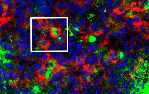
Splenic dendritic cells (green) phagocytize erythrocytes infected by Plasmodium chabaudi (red) in the red pulp of the spleen (image: Henrique Borges da Silva)
A study shows that cells present in splenic red pulp can control and eliminate the parasite that causes malaria in the first few days of infection.
A study shows that cells present in splenic red pulp can control and eliminate the parasite that causes malaria in the first few days of infection.
Splenic dendritic cells (green) phagocytize erythrocytes infected by Plasmodium chabaudi (red) in the red pulp of the spleen (image: Henrique Borges da Silva)
By Elton Alisson
Agência FAPESP – Researchers at the University of São Paulo’s Biomedical Science Institute (ICB-USP) have elucidated one of the mechanisms used by the immune system to control the presence of plasmodia in the host’s blood stream. Plasmodia are protozoan parasites of the genus Plasmodium, five species of which cause malaria.
The research was performed in collaboration with colleagues at USP’s Chemistry Institute, the Gulbenkian Science Institute (IGC) in Portugal, the MRC National Institute for Medical Research in the United Kingdom, the Federal University of São Paulo (UNIFESP) in Brazil, and the University of Orléans in France.
The researchers discovered that dendritic cells present in large numbers in the red pulp of the spleen (a major site for the control of bloodborne infections such as malaria) are activated to contain and eliminate plasmodia from the blood in the first few days of infection by means of phagocytosis of infected red blood cells (erythrocytes). Phagocytosis is the process by which cells called phagocytes ingest or engulf other cells or particles.
The discovery resulted from the PhD research project “Characterization of effector mechanisms of innate and acquired immunity in a chronic malaria model of CD28KO mice infected with Plasmodium chabaudi AS,” supported by a scholarship from FAPESP. The findings were described in an article published in the journal PLoS Pathogens.
“We found that dendritic cells begin phagocytizing infected erythrocytes shortly after infection by the parasite and play a very important role in limiting initial parasitemia, the number of parasites present in the blood stream. They’re also crucial to activation of the immune response against these pathogens,” said Henrique Borges da Silva, a postdoctoral researcher at ICB-USP with a scholarship from FAPESP and first author of the study.
According to Silva, the splenic red pulp was already known to be a region of the organism that specializes in controlling bloodborne infections such as malaria, as it contains large numbers of macrophages, white blood cells with a high capacity to phagocytize old or infected cells, as well as certain types of pathogens (microorganisms that cause disease).
However, it was not known how other phagocytic cells in this region of the spleen contribute specifically to the elimination of pathogens from the blood, including the plasmodia that cause malaria, Silva explained.
Given the large population of dendritic cells in the splenic red pulp, the researchers raised the hypothesis that dendritic cells may play this role by phagocytizing plasmodium-infected erythrocytes.
To test this hypothesis, the investigators intravenously inoculated C57BL6.CD11c-YFP transgenic mice, whose splenic dendritic cells express yellow fluorescent protein (YFP), with a transgenic fluorescent strain of P. chabaudi.
At various times after infection, they analyzed interactions between erythrocytes infected with the transgenic fluorescent parasite and splenic dendritic cells. They used a confocal intravital microscope, which can detect fluorescence at varying depths and produces 3D images of several layers of tissue, enabling scientists to observe entire organs in live organisms.
The experiments were performed partly at ICB-USP’s Research Support Facilities Center (CEFAP), one of the projects funded by FAPESP’s Multi-User Equipment Program. Analysis of the 3D images produced using this technique showed that the animals’ splenic dendritic cells recognized and actively phagocytized infected erythrocytes during the acute phase of blood infection by Plasmodium.
In this phase, which in mice starts on the first day after infection, before the appearance of symptoms of the disease, part of the population of splenic dendritic cells migrated to regions rich in T-lymphocytes and stimulated the action of CD4+ T cells, which are extremely important to the immune system, activating the host’s immune response to infection.
“It’s the first time anyone has demonstrated directly in vivo [in live organisms] and in situ [in a viable organ] the interactions between dendritic cells and infected erythrocytes and such a high level of phagocytosis by the cells of the splenic immune system,” Silva told Agência FAPESP.
According to Silva, “Previous studies carried out to evaluate phagocytosis of infected erythrocytes by other kinds of cells in the splenic immune system using other techniques, such as flow cytometry, reported very low percentages, which were not consistent with the level of parasitemia we observed in plasmodium-infected mice or with the spleen’s importance in eliminating plasmodia as reported hitherto.”
Control of parasitemia
To determine whether parasite elimination by splenic dendritic cell phagocytosis helped to control parasitemia and increase the survival rate for infected mice, the researchers performed a second set of experiments, in which they first inoculated CD11c-DTR mice with human diphtheria toxin (DTx). Silva exlained that DTx induced the elimination of dendritic cells in these transgenic mice. “When we treated these mice with human diphtheria toxin, all of their splenic dendritic cells were eliminated.”
After treating the mice with DTx, the researchers infected them with P. chabaudi to gauge the role of splenic dendritic cells in the evolution of the infection. They observed that the mice lacking splenic dendritic cells displayed higher levels of parasitemia and mortality due to infection by the parasite than mice that had not been injected with DTx.
“The results showed that splenic dendritic cells, at least in our experimental malaria model, play a key role in containing the acute phase of infection by plasmodia,” Silva said.
The researchers also found that in the first two days after infection the transgenic mice injected with DTx displayed high levels of parasitemia compared with those not injected with DTx.
A possible explanation for such high levels of parasitemia in the acute phase of the infection was that the absence of splenic dendritic cells impaired direct elimination of plasmodium-infected erythrocytes.
“The much higher levels of parasitemia in early-stage infection mice lacking splenic dendritic cells suggests that the phagocytic role of these cells may be an important means of containing the number of parasites in the blood stream,” Silva said.
The researchers also observed that splenic dendritic cells appeared to reach full maturity only when parasitemia had peaked and been controlled, which occurred at the end of the acute infectious phase.
For example, only when their maturation is complete can splenic dendritic cells stimulate production of memory-specific CD4+ T cells, which are fundamental to the organism’s response to malaria and its capacity to contain new infections by plasmodia.
“This discovery may be useful for the development or enhancement of immunization strategies, especially in the blood stage of plasmodium infection, when the main immune responses against the parasite take place,” Silva said.
One of the researchers’ next steps, he added, will be to investigate how splenic dendritic cells recognize and phagocytize infected erythrocytes.
The article “In vivo approaches reveal a key role for DCs in CD4+ T cell activation and parasite clearance during the acute phase of experimental blood-stage malaria” (doi: 10.1371/journal.ppat.1004598), by Silva et al., can be read in the journal PLoS Pathogens at http://journals.plos.org/plospathogens/article?id=10.1371/journal.ppat.1004598.
The Agency FAPESP licenses news via Creative Commons (CC-BY-NC-ND) so that they can be republished free of charge and in a simple way by other digital or printed vehicles. Agência FAPESP must be credited as the source of the content being republished and the name of the reporter (if any) must be attributed. Using the HMTL button below allows compliance with these rules, detailed in Digital Republishing Policy FAPESP.
