

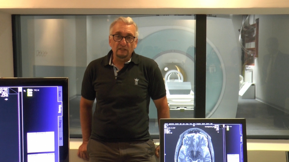
Researcher Paulo Saldiva with the Magnetom 7T MRI in the background (photo: Leandro Negro)
Equipped with Latin America's first 7-Tesla whole-body magnetic resonance imaging scanner, the lab will study cadavers and try to make autopsies less invasive.
Equipped with Latin America's first 7-Tesla whole-body magnetic resonance imaging scanner, the lab will study cadavers and try to make autopsies less invasive.
Researcher Paulo Saldiva with the Magnetom 7T MRI in the background (photo: Leandro Negro)
By Elton Alisson
Agência FAPESP – On March 13, the University of São Paulo’s School of Medicine (FM-USP) unveiled an advanced research facility called the Autopsy Room Imaging Platform, whose Portuguese-language acronym is PISA. The new facility’s centerpiece is a Magnetom 7T MRI, Latin America’s first 7-Tesla whole-body magnetic resonance imaging scanner.
Installed in a 500 sq. m. underground bunker in a lot adjacent to the School of Medicine’s main complex, the laboratory also contains computed tomography (CT), ultrasound and X-ray imaging equipment.
The Magnetom cost US$7.695 million (about R$24 million). Its purchase was funded by three institutions: FAPESP, under the project “Imaging platform in the autopsy room” and the Multi-User Equipment Program (EMU); the University of São Paulo (USP); and the São Paulo State Department of Health, through Fundação Faculdade de Medicina.
“The facility will advance knowledge of new diseases and contribute to research in several fields, with a significant impact on health, as well as helping to enhance medical education and training,” said FAPESP President Celso Lafer during the ceremony held to unveil PISA.
Made in Germany and the United Kingdom, the 7-Tesla scanner is capable of simultaneously producing several ultra-high resolution whole-body images. Similar machines are in service elsewhere, such as in Europe, the United States and Japan, but they are smaller and are not whole-body scanners, according to Edson Amaro Júnior, a professor of radiology at FM-USP.
“There are about seventy 7T MRI scanners in the northern hemisphere and one in Australia, but most are very small and are mainly used experimentally,” he said. Another difference is that the Brazilian machine will be used to examine cadavers received by São Paulo City’s Death Certification Service (SVOC). Run by USP, this is the largest service of its kind in the world, performing some 14,000 natural-death autopsies per year.
Many more post-mortem case studies of diseases will now be feasible, and the imaging equipment’s diagnostic accuracy will be validated with greater precision, according to researchers who are participating in the project.
“A better understanding of natural-death causes will enable us to develop new treatments and contribute to improvements in the quality of the healthcare provided to patients, because the prime aim of hospital care quality control is to ensure that the patient enters and leaves the hospital alive,” said Paulo Hilário Saldiva, Full Professor of Pathology at FM-USP.
“If the patient dies, the questions that must be asked are whether everything possible was done, whether the diagnostic hypotheses were correct, and whether complications arose during treatment. This is extremely important to the formulation of a hospital quality policy.”
The 7T MRI is so powerful that the images it produces are 5.4 times larger than those produced by the 3T scanners used in some Brazilian hospitals and show submillimeter anatomical details as fine as a human hair.
Brain imaging
The first images produced by the Magnetom will be of the brain, as the machine is factory-set to analyze only that organ.
The researchers at FM-USP, however, are working on devices that will enable the scanner to image other organs. “We have a postdoc who spent a few years in the US learning how to build such devices,” Amaro said.
The researchers will also help to evaluate and develop applications of 7T MRI scanners for live patients.
Safety precautions explain why the machine has not yet been approved for clinical use, given its powerful magnetic field. If the machine’s magnet, which weighs three tons and is always energized, were attached to a crane, it could lift the equivalent of 20 mid-size cars, according to Amaro.
Furthermore, the radiofrequency (RF) waves it emits may heat all parts of the patient’s body, whereas in clinical applications heating should ideally be confined to the body part being analyzed.
“We’re going to use optical thermometers to measure temperature in various areas of the body and find out which ones the scanner heats. Any potential side effects will be carefully identified,” Amaro said.
“We’ll have the opportunity to help evaluate and develop applications of the 7T for live patients,” said Guido Cerri, a professor at FM-USP’s Radiology Department and a former São Paulo State health secretary.
Comparing autopsies
The researchers at PISA are already working on a project, called the Brazilian Imaging and Autopsy Study (BIAS), that calls for the execution of 1,000 minimally invasive prospective autopsies for comparison with conventional autopsies.
So far, they have performed some 900 autopsies using CT, ultrasound and X-ray imaging, according to Saldiva.
He said that the number of natural-death autopsies is falling worldwide, partly owing to a rising trend in families refusing to consent to the procedure. However, Saldiva pointed out, autopsy “still occupies a unique space in medicine and can help elucidate the mechanisms of emerging diseases, such as arteriosclerosis or Alzheimer’s, which haven’t benefited from advances in molecular biology because you can’t take a biopsy from a living person’s brain or heart.”
To facilitate acceptance of the procedure and to persuade family members to help to determine the cause of a relative’s natural death, researchers at FM-USP are working on the “The Use of Modern Autopsy Techniques to Investigate Human Diseases” Thematic Project, led by Saldiva. The aim is to develop diagnostic imaging techniques that can be used to make autopsies less invasive than the conventional procedure.
The project is supported by FAPESP and has been given an additional boost by the unveiling of PISA.
“The concept that we’re working on to persuade families to consent to autopsy is knowledge donation,” Saldiva said.
More than half of all deaths worldwide are from unknown causes or very vaguely defined causes without utility for comparative studies, such as cardiac arrest, a feature of all deaths, he added.
“If we can increase the number of natural-death autopsies by using minimally invasive techniques developed in the lab, we can achieve better classification of the causes of death,” he said. “We have a rare opportunity to obtain high-resolution images and at the same time collect data on the anatomical and cellular aspects of organs.”
Read more about the University of São Paulo’s Autopsy Room Imaging Platform (in Portuguese) at agencia.fapesp.br/a_morte_explica_a_vida/20801/.
The Agency FAPESP licenses news via Creative Commons (CC-BY-NC-ND) so that they can be republished free of charge and in a simple way by other digital or printed vehicles. Agência FAPESP must be credited as the source of the content being republished and the name of the reporter (if any) must be attributed. Using the HMTL button below allows compliance with these rules, detailed in Digital Republishing Policy FAPESP.
